
Page 1218 - Equine Clinical Medicine, Surgery and Reproduction, 2nd Edition
P. 1218
Eyes 1193
VetBooks.ir the most frequent congenital ocular defect in foals. 11.105
Very small incipient lens opacities are common
and not associated with blindness. As the cataracts
mature and become opaque the degree of blindness
increases. Most veterinary ophthalmologists recom-
mend surgical removal of cataracts in foals less than
6 months of age if the foal is healthy, no uveitis or
other ocular problems are present, and the animal’s
personality will allow them to tolerate aggressive
topical medical therapy.
Aetiology/pathophysiology
The majority of adult equine cataracts are acquired,
with chronic uveitis being the most common cause
(see Table 11.5). The lens is nourished by the aqueous
humour and any alteration in its production, compo-
sition or flow can have adverse effects on lens metab-
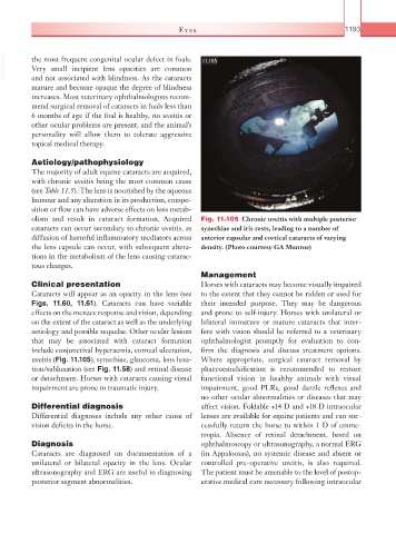
olism and result in cataract formation. Acquired Fig. 11.105 Chronic uveitis with multiple posterior
cataracts can occur secondary to chronic uveitis, as synechiae and iris rests, leading to a number of
diffusion of harmful inflammatory mediators across anterior capsular and cortical cataracts of varying
the lens capsule can occur, with subsequent altera- density. (Photo courtesy GA Munroe)
tions in the metabolism of the lens causing catarac-
tous changes.
Management
Clinical presentation Horses with cataracts may become visually impaired
Cataracts will appear as an opacity in the lens (see to the extent that they cannot be ridden or used for
Figs. 11.60, 11.61). Cataracts can have variable their intended purpose. They may be dangerous
effects on the menace response and vision, depending and prone to self-injury. Horses with unilateral or
on the extent of the cataract as well as the underlying bilateral immature or mature cataracts that inter-
aetiology and possible sequelae. Other ocular lesions fere with vision should be referred to a veterinary
that may be associated with cataract formation ophthalmologist promptly for evaluation to con-
include conjunctival hyperaemia, corneal ulceration, firm the diagnosis and discuss treatment options.
uveitis (Fig. 11.105), synechiae, glaucoma, lens luxa- Where appropriate, surgical cataract removal by
tion/subluxation (see Fig. 11.58) and retinal disease phaecoemulsification is recommended to restore
or detachment. Horses with cataracts causing visual functional vision in healthy animals with visual
impairment are prone to traumatic injury. impairment, good PLRs, good dazzle reflexes and
no other ocular abnormalities or diseases that may
Differential diagnosis affect vision. Foldable +14 D and +18 D intraocular
Differential diagnoses include any other cause of lenses are available for equine patients and can suc-
vision deficits in the horse. cessfully return the horse to within 1 D of emme-
tropia. Absence of retinal detachment, based on
Diagnosis ophthalmoscopy or ultrasonography, a normal ERG
Cataracts are diagnosed on documentation of a (in Appaloosas), no systemic disease and absent or
unilateral or bilateral opacity in the lens. Ocular controlled pre- operative uveitis, is also required.
ultrasonography and ERG are useful in diagnosing The patient must be amenable to the level of postop-
posterior segment abnormalities. erative medical care necessary following intraocular