
Page 1222 - Equine Clinical Medicine, Surgery and Reproduction, 2nd Edition
P. 1222
Eyes 1197
VetBooks.ir dorsal thorax. These pre-ganglionic sympathetic 11.108
axons then travel through the cervicothoracic and
middle cervical ganglia and continue up the neck, in
the vagosympathetic trunk, to synapse in the cranial
cervical ganglion. The post-ganglionic sympathetic
axons then continue forwards, where they pass
through the middle ear, join the ophthalmic branch
of the trigeminal nerve and distribute to the sweat
glands of the head, smooth muscles of the periorbita
and eyelids, and the iris dilator muscle.
Lesions causing cranial sympathetic denervation,
and thus leading to Horner’s syndrome, can occur
anywhere along this pathway. Loss of sympathetic
innervation to Muller’s muscle of the upper eyelid
and tissue of the lower eyelid results in narrowing of
the palpebral fissure and ptosis (drooping of the upper
eyelid). Ipsilateral facial sweating and regional hyper-
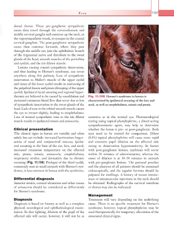
thermia are believed to be caused by vasodilation and Fig. 11.108 Horner’s syndrome in horses is
increased cutaneous blood flow that occur due to loss characterised by ipsilateral sweating of the face and
of sympathetic innervation to the sweat glands of the neck, as well as enophthalmos, miosis and ptosis.
head. Lack of tone in the orbital smooth muscle causes
the eye to retract slightly, leading to enophthalmos.
Loss of normal sympathetic tone to the iris dilator extensive as in the normal eye. Pharmacological
muscle results in ipsilateral miosis and anisocoria. testing using topical phenylephrine, a direct-acting
sympathomimetic agent, may help to determine
Clinical presentation whether the lesion is pre- or post-ganglionic. Both
The clinical signs in horses are variable and often eyes need to be treated for comparison. Dilute
subtle but can include: increased lacrimation; hyper- (0.1%) topical phenylephrine will cause more rapid
aemia of nasal and conjunctival mucosa; ipsilat- and extensive pupil dilation on the affected side
eral sweating at the base of the ear, face, and neck; owing to denervation hypersensitivity. In horses
increased cutaneous temperature on the affected with post-ganglionic lesions, mydriasis will occur
side; ptosis; miosis; anisocoria; enophthalmos; within 20 minutes of administration, whereas the
inspiratory stridor; and dermatitis due to chronic onset of dilation is at 30–50 minutes in animals
sweating (Fig. 11.108). Prolapse of the third eyelid, with pre- ganglionic lesions. The guttural pouches
commonly seen in small animals with Horner’s syn- and the pharynx of all patients should be examined
drome, is less common in horses with the syndrome. endoscopically, and the jugular furrows should be
palpated for swellings. A history of recent intrave-
Differential diagnosis nous or intramuscular injections in the neck should
Anterior uveitis, corneal ulceration and other causes be obtained. Radiographs of the cervical vertebrae
of anisocoria should be considered as differentials or thorax may also be indicated.
for Horner’s syndrome.
Management
Diagnosis Treatment will vary depending on the underlying
Diagnosis is based on history as well as a complete cause. There is no specific treatment for Horner’s
physical, neurological and ophthalmological exami- syndrome; however, topical phenylephrine may be
nation. In dim lighting, dilation of the pupil of the used therapeutically for temporary alleviation of the
affected side will occur; however, it will not be as associated clinical signs.