
Page 1221 - Equine Clinical Medicine, Surgery and Reproduction, 2nd Edition
P. 1221
1196 CHAPTER 11
VetBooks.ir 1 mg/kg p/o q12 h; methazolamide, 0.25 mg/kg p/o and haemorrhage, and decreased vision or blindness.
Many eyes need to be treated again in 6–12 months.
q24 h), but potassium supplementation is typically
required. Topical and systemic anti-inflammatory
some clinicians suggest that it should be reserved for
medications should be used initially to help con- Cyclocryoablation has also been used; however,
trol intraocular inflammation and increase patient use in blind eyes only. Again, any decrease in aque-
comfort. ous production may be only temporary.
Laser cyclophotoablation can be used to decrease Surgical techniques to increase aqueous outflow
the amount of aqueous humour produced by the cili- (e.g. gonioimplants, sclerostomies and iridectomies)
ary body in eyes with the potential for vision that do have also been used in horses with glaucoma with
not respond to antiglaucoma medications. Existing varying success. Horses with glaucoma should have
intraocular inflammation must be controlled prior their IOP measured regularly in order to monitor the
to treatment and other intraocular diseases such as response to therapy. Often, affected eyes will become
neoplasia should be ruled out. If corneal ulcers are blind and chronically painful. Enucleation or evis-
present, they should be treated prior to laser surgery. ceration with intrascleral prosthesis is the treatment
Systemic anti-inflammatory medications are required of choice in these cases. Where primary glaucoma is
for 7–10 days following laser therapy. Lasers appear suspected, repeated measurements of the IOP should
to be very effective at controlling IOP and helping be taken in the fellow (predisposed) eye 3–4 times per
to preserve vision, with over 50% of eyes remain- year for life or until the eye becomes glaucomatous.
ing sighted. However, antiglaucoma medications
are usually required indefinitely after surgery. Post- Prognosis
surgical complications include continued or recur- The prognosis for vision is guarded. The most effec-
ring elevation in IOP, hypotony, blepharoedema, tive long-term therapy currently available appears to
chemosis, corneal oedema, ocular haemorrhage, be cyclophotoablation in combination with topical
corneal ulceration, cataract formation, vitreal fibrin antiglaucoma medications.
NEUROLOGIC DISORDERS OF THE EYE
HORNER’S SYNDROME
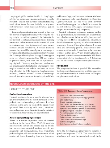
Table 11.10 Causes of Horner’s syndrome
Definition/overview • Severe head, neck and chest trauma
Horner’s syndrome is not a specific disease, but a • Cranial thoracic neoplasia/space-occupying masses
syndrome that involves the loss or disruption of sym- • Otitis media/interna
pathetic innervation to the eye and adnexa. It is char- • Cervical neoplasia or abscesses
acterised in the horse by ptosis of the upper eyelid, • Drug injection into the carotid artery or jugular vein
ipsilateral facial sweating, mild miosis, enophthal- • Guttural pouch disease or surgery (e.g. carotid artery
ligation for facial surgery or guttural pouch epistaxis)
mos and regional hyperthermia. It may be unilateral • Oesophageal rupture, obstruction or surgery
or bilateral and may or may not be permanent. • Periorbital abscesses or tumours
• Post-anaesthetic myopathy
Aetiology/pathophysiology • Equine protozoal meningoencephalitis
There are a number of possible causes of Horner’s • Cauda equine neuritis/polyneuritis equi
syndrome in the horse (Table 11.10). Sympathetic • Systemic aspergillosis
• Central nervous system infection or neoplasia
innervation to the eye and adnexa may be divided
into three neuroanatomical sections: central, pre-
ganglionic and post-ganglionic. The sympathetic down the tectotegmentospinal tract to synapse at
pathway begins with the central component, which spinal cord segments T1–T3. The axons leave the
consists of fibres descending from the brainstem, spinal cord and enter the sympathetic trunk in the