
Page 1220 - Equine Clinical Medicine, Surgery and Reproduction, 2nd Edition
P. 1220
Eyes 1195
VetBooks.ir 11.106 11.107
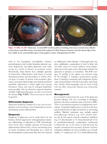
Figs. 11.106, 11.107 Glaucoma. A raised IOP results in globe stretching and creates corneal striae (Haab’s
striae), linear track-like lesions associated with oedema (11.106). Posterior luxation with the dorsal edge of the
lens visible in the ventromedial aspect of the pupil in a horse with glaucoma (11.107).
such as iris hypoplasia, microphakia, cataract, to traditional uveitis therapy. A thorough and com-
goniodysgenesis and retinal dysplasia (anterior seg- plete ophthalmic examination is vital to help rule
ment dysgenesis). Secondary glaucoma most often out other causes of corneal oedema, vision impair-
occurs as a result of chronic or recurrent uveitis. ment and ocular pain, and to determine whether the
Historically, these horses have multiple episodes glaucoma is primary or secondary. The IOP aver-
of intraocular inflammation with bouts of ocular ages 24 mmHg in the equine eye (normal range
cloudiness/oedema and discomfort, as well as clini- 15–30 mmHg). A Tonopen measurement greater
cal signs of uveitis. In horses with secondary glau- than 35 mmHg is consistent with a diagnosis of glau-
coma, associated clinical signs may include posterior coma. Examination of the ICA may show abnormal-
synechiae (adhesions), a miotic pupil and cataract ities. Ocular ultrasonography may be used to help
formation. These eyes may be enlarged (buphthal- rule out other intraocular diseases (e.g. intraocular
mos), possibly with an ulcerative exposure keratitis, tumour).
and lens subluxation/luxation can also occur late in
the disease (Fig. 11.107). These eyes may or may not Management
be painful. It is essential to determine the cause of the glaucoma
because therapy will vary according to aetiology;
Differential diagnosis however, the most common cause in horses is ERU.
Glaucoma should be considered in any case of unex- There is inconsistent response to antiglaucoma medi-
plained corneal oedema, vision impairment or severe cations. Treatment of glaucoma is centred on decreas-
unrelenting ocular inflammation. ing the production of aqueous humour or increasing
outflow. Medical treatment may include topical
Diagnosis beta-adrenergic blockers (e.g. 0.5% timolol male-
Diagnosis of glaucoma can be made based on the ate q12 h) and topical carbonic anhydrase inhibitors
history, clinical appearance and applanation tonom- (e.g. 2% dorzolamide or 1% brinzolamide q8–12 h).
etry illustrating an elevation in IOP. Historically, A timolol/dorzolamide combination medication
these horses have multiple episodes of intraocular is offered for use in the horse to help decrease the
inflammation followed by a severe unrelenting bout number of medications necessary. Systemic carbonic
of ocular cloudiness and discomfort (as a result of anhydrase inhibitors are also available (e.g. acetazol-
the development of glaucoma) that does not respond amide, 2–3 mg/kg p/o q6–12 h; dichlorphenamide,