
Page 1225 - Equine Clinical Medicine, Surgery and Reproduction, 2nd Edition
P. 1225
1200 CHAPTER 11
VetBooks.ir 11.109 retinal pigmented epithelial cell congestion with
ceroid-lipofuscin. Ceroid-lipofuscin deposition is
also always observed in the endothelial capillaries in
the spinal cord and, occasionally, in the liver and GI
tract.
Management
Treatment for horses with EMND involves dietary
supplementation with vitamin E and access to pas-
ture or fresh forage.
Prognosis
The prognosis is variable. In 40% of cases, marked
improvement in clinical signs is seen within
4–6 weeks after relocation to another stable and/
or administration of dietary antioxidants. However,
40% of horses are euthanased or die owing to
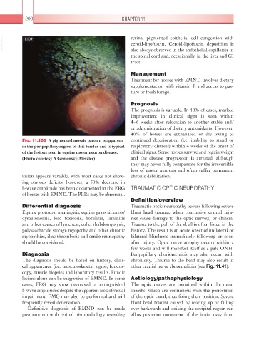
Fig. 11.109 A pigmented mosaic pattern is apparent continued deterioration (i.e. inability to stand or
in the peripapillary region of this fundus and is typical respiratory distress) within 4 weeks of the onset of
of the lesions seen in equine motor neuron disease. clinical signs. Some horses survive and regain weight
(Photo courtesy A Gemensky-Metzler) and the disease progression is arrested, although
they may never fully compensate for the irreversible
loss of motor neurons and often suffer permanent
vision appears variable, with most cases not show- chronic debilitation.
ing obvious deficits; however, a 50% decrease in
b-wave amplitude has been documented in the ERG TRAUMATIC OPTIC NEUROPATHY
of horses with EMND. The PLRs may be abnormal.
Definition/overview
Differential diagnosis Traumatic optic neuropathy occurs following severe
Equine protozoal meningitis, equine grass sickness/ blunt head trauma, when concussive cranial inju-
dysautonomia, lead toxicosis, botulism, laminitis ries cause damage to the optic nerve(s) or chiasm.
and other causes of lameness, colic, rhabdomyolysis, Trauma to the poll of the skull is often listed in the
polysaccharide storage myopathy and other chronic history. The result is an acute onset of unilateral or
myopathies, iliac thrombosis and senile retinopathy bilateral blindness immediately following or soon
should be considered. after injury. Optic nerve atrophy occurs within a
few weeks and will manifest itself as a pale ONH.
Diagnosis Peripapillary chorioretinitis may also occur with
The diagnosis should be based on history, clini- chronicity. Trauma to the head may also result in
cal appearance (i.e. musculoskeletal signs), fundos- other cranial nerve abnormalities (see Fig. 11.41).
copy, muscle biopsies and laboratory results. Fundic
lesions alone can be suggestive of EMND. In some Aetiology/pathophysiology
cases, ERG may show decreased or extinguished The optic nerves are contained within the dural
b-wave amplitudes despite the apparent lack of visual sheaths, which are continuous with the periosteum
impairment. EMG may also be performed and will of the optic canal, thus fixing their position. Severe
frequently reveal denervation. blunt head trauma caused by rearing up or falling
Definitive diagnosis of EMND can be made over backwards and striking the occipital region can
post mortem with retinal histopathology revealing allow posterior movement of the brain away from