
Page 1230 - Equine Clinical Medicine, Surgery and Reproduction, 2nd Edition
P. 1230
Eyes 1205
VetBooks.ir 11.116 Aetiology/pathophysiology
Habronemiasis is caused by the aberrant migration
of nematode larvae of the species Habronema mus-
cae, H. microstoma and Draschia megastoma. The adult
parasite resides in the stomach of the horse. The
eggs or larvae are passed in the faeces and ingested
by the larvae of the intermediate host (either the
house fly Musca domestica or the stable fly Stomoxys
calcitrans). Horses are infected following ingestion
of an infected adult fly, or infectious L3 larvae may
be deposited on wounds around the eye or near the
mouth. Larval migration through tissue incites a
granulomatous inflammatory response, which can
become walled off into discrete nodules or multi-
lobulated masses that become caseous and necrotic.
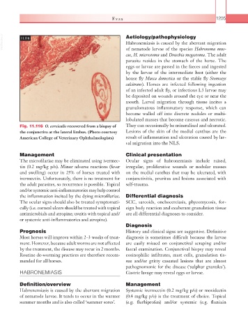
Fig. 11.116 O. cervicalis recovered from a biopsy of They can occasionally be mineralised and ulcerated.
the conjunctiva at the lateral limbus. (Photo courtesy Lesions of the skin of the medial canthus are the
American College of Veterinary Ophthalmologists) result of inflammation and ulceration caused by lar-
val migration into the NLS.
Management Clinical presentation
The microfilariae may be eliminated using ivermec- Ocular signs of habronemiasis include raised,
tin (0.2 mg/kg p/o). Minor adverse reactions (fever irregular, proliferative wounds or nodular masses
and swelling) occur in 25% of horses treated with on the medial canthus that may be ulcerated, with
ivermectin. Unfortunately, there is no treatment for conjunctivitis, pruritus and lesions associated with
the adult parasites, so recurrence is possible. Topical self-trauma.
and/or systemic anti-inflammatories may help control
the inflammation incited by the dying microfilariae. Differential diagnosis
The ocular signs should also be treated symptomati- SCC, sarcoids, onchocerciasis, phycomycosis, for-
cally (i.e. corneal ulcers should be treated with topical eign body reaction and exuberant granulation tissue
antimicrobials and atropine; uveitis with topical and/ are all differential diagnoses to consider.
or systemic anti-inflammatories and atropine).
Diagnosis
Prognosis History and clinical signs are suggestive. Definitive
Most horses will improve within 2–3 weeks of treat- diagnosis is sometimes difficult because the larvae
ment. However, because adult worms are not affected are easily missed on conjunctival scraping and/or
by the treatment, the disease may recur in 2 months. faecal examination. Conjunctival biopsy may reveal
Routine de-worming practices are therefore recom- eosinophilic infiltrates, mast cells, granulation tis-
mended for all horses. sue and/or gritty caseated lesions that are almost
pathognomonic for the disease (‘sulphur granules’).
HABRONEMIASIS Gastric lavage may reveal eggs or larvae.
Definition/overview Management
Habronemiasis is caused by the aberrant migration Systemic ivermectin (0.2 mg/kg p/o) or moxidectin
of nematode larvae. It tends to occur in the warmer (0.4 mg/kg p/o) is the treatment of choice. Topical
summer months and is also called ‘summer sores’. (e.g. flurbiprofen) and/or systemic (e.g. flunixin