
Page 1226 - Equine Clinical Medicine, Surgery and Reproduction, 2nd Edition
P. 1226
Eyes 1201
VetBooks.ir the fixed intracanalicular portion of the optic nerves. 11.110
This may cause stretching, shearing and/or avulsion
of the retinal ganglion cell axons/optic nerve(s) or
chiasm, resulting in optic nerve atrophy and sudden
blindness. Partial or complete visual loss occurs in
the affected eye(s) within 24 hours of injury.
Clinical presentation
Horses with traumatic optic neuropathy present
with a history of sudden onset of blindness with or
without a known history of trauma. The pupil(s) is
(are) fixed and dilated with sluggish to absent PLRs
in the affected eye(s). Ophthalmic lesions are not
usually seen initially because of the often retrobul-
bar nature of the injury.
Ophthalmoscopic lesions, including peripapillary
and/or ONH oedema or haemorrhage and exudation
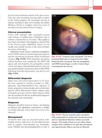
into the vitreous may be present within 24–48 hours Fig. 11.110 Traumatic optic neuropathy. This horse
of injury (Fig. 11.110). With chronicity, the lamina presented blind and was suspected to have fallen
cribrosa becomes more prominent, the ONH will backwards after rearing up. Note the peripapillary
appear pale and atrophied, the peripapillary retinal and ONH haemorrhages, as well as haemorrhagic
vessels will appear diminished/attenuated, and focal streaming into the vitreous.
grey patches medial, lateral and ventral to the ONH,
indicating choroidal degeneration, may also be seen
(Fig. 11.111). 11.111
Differential diagnosis
Optic nerve and retinal degeneration in the horse
has been reported to develop secondary to ERU,
glaucoma, hypovolaemia/blood loss, exposure to
toxins, progressive retinal atrophy and carotid artery
ligation. Other differentials include exudative optic
neuropathy and optic neuritis. Brain injuries should
also be considered in the list of differential diagnoses
for traumatic optic neuropathy.
Diagnosis
Diagnosis should be based on history and findings
on physical and ophthalmic examination. A failure
to navigate photopic and scotopic maze tests is also
observed.
Fig. 11.111 Chronic traumatic optic neuropathy.
Management This yearling fell over backwards 2 months previously
Treatment when cases are presented acutely tradi- and was immediately blind but without any fundic
tionally involves high doses of anti-inflammatories changes. The fundus of both eyes now shows classic
such as systemic corticosteroids (e.g. dexametha- changes of the condition in the optic disc and tapetal/
sone), NSAIDs and dimethylsulphoxide in order to non-tapetal fundus. (Photo courtesy GA Munroe)