
Page 1229 - Equine Clinical Medicine, Surgery and Reproduction, 2nd Edition
P. 1229
1204 CHAPTER 11
VetBooks.ir PARASITIC DISEASES OF THE EYE
ONCHOCERCIASIS
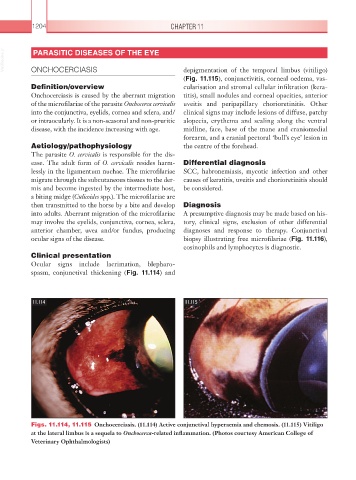
depigmentation of the temporal limbus (vitiligo)
(Fig. 11.115), conjunctivitis, corneal oedema, vas-
Definition/overview cularisation and stromal cellular infiltration (kera-
Onchocerciasis is caused by the aberrant migration titis), small nodules and corneal opacities, anterior
of the microfilariae of the parasite Onchocerca cervicalis uveitis and peripapillary chorioretinitis. Other
into the conjunctiva, eyelids, cornea and sclera, and/ clinical signs may include lesions of diffuse, patchy
or intraocularly. It is a non-seasonal and non-pruritic alopecia, erythema and scaling along the ventral
disease, with the incidence increasing with age. midline, face, base of the mane and craniomedial
forearm, and a cranial pectoral ‘bull’s eye’ lesion in
Aetiology/pathophysiology the centre of the forehead.
The parasite O. cervicalis is responsible for the dis-
ease. The adult form of O. cervicalis resides harm- Differential diagnosis
lessly in the ligamentum nuchae. The microfilariae SCC, habronemiasis, mycotic infection and other
migrate through the subcutaneous tissues to the der- causes of keratitis, uveitis and chorioretinitis should
mis and become ingested by the intermediate host, be considered.
a biting midge (Culicoides spp.). The microfilariae are
then transmitted to the horse by a bite and develop Diagnosis
into adults. Aberrant migration of the microfilariae A presumptive diagnosis may be made based on his-
may involve the eyelids, conjunctiva, cornea, sclera, tory, clinical signs, exclusion of other differential
anterior chamber, uvea and/or fundus, producing diagnoses and response to therapy. Conjunctival
ocular signs of the disease. biopsy illustrating free microfilariae (Fig. 11.116),
eosinophils and lymphocytes is diagnostic.
Clinical presentation
Ocular signs include lacrimation, blepharo-
spasm, conjunctival thickening (Fig. 11.114) and
11.114 11.115
Figs. 11.114, 11.115 Onchocerciasis. (11.114) Active conjunctival hyperaemia and chemosis. (11.115) Vitiligo
at the lateral limbus is a sequela to Onchocerca-related inflammation. (Photos courtesy American College of
Veterinary Ophthalmologists)