
Page 1228 - Equine Clinical Medicine, Surgery and Reproduction, 2nd Edition
P. 1228
Eyes 1203
VetBooks.ir 11.112 11.113
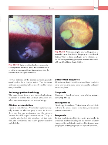
Fig. 11.113 Proliferative optic neuropathy present at
8–9 o’clock was identified in this pony as an incidental
finding. There is also a small optic nerve coloboma at
the 6 o’clock position (typical) that was not associated
with any identifiable visual deficits.
Fig. 11.112 Optic neuritis of unknown cause in
a young Welsh Section A pony. Note the exudation
of white–cream material and haemorrhage into the
vitreous from the optic nerve head.
obscure portions of the retina) and it is generally Differential diagnosis
considered to be a benign lesion. This incidental This disease should be differentiated from exudative
finding is seen unilaterally, primarily in older horses optic neuritis, traumatic optic neuropathy and optic
(>15 years old). nerve neoplasia.
Aetiology/pathophysiology Diagnosis
The cause is not known, and the pathophysiology Diagnosis is based on history and clinical appear-
is unclear. The mass has a similar appearance to a ance (Fig. 11.113).
schwannoma/astrocytoma on histopathology.
Management
Clinical presentation No therapy is available. Vision is not affected clini-
Vision is not affected. Proliferative optic neurop- cally and the lesions appear to be stable, so treatment
athy is seen as white or grey masses on or near appears unnecessary.
the optic disc and protruding into the vitreous
humour in middle-aged or older horses. They are Prognosis
typically attached at the periphery of the optic Benign exudative/proliferative optic neuropathy is
disc, are vascularised and can be pedunculated or usually an incidental finding. In the absence of other
multilobular. changes, this condition is considered benign and non-
progressive and the prognosis for vision is excellent.