
Page 1323 - Equine Clinical Medicine, Surgery and Reproduction, 2nd Edition
P. 1323
1298 CHAPTER 13
VetBooks.ir 13.3 13.4
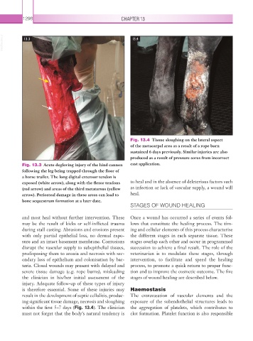
Fig. 13.4 Tissue sloughing on the lateral aspect
of the metacarpal area as a result of a rope burn
sustained 6 days previously. Similar injuries are also
produced as a result of pressure sores from incorrect
Fig. 13.3 Acute degloving injury of the hind cannon cast application.
following the leg being trapped through the floor of
a horse trailer. The long digital extensor tendon is
exposed (white arrow), along with the flexor tendons to heal and in the absence of deleterious factors such
(red arrow) and areas of the third metatarsus (yellow as infection or lack of vascular supply, a wound will
arrow). Periosteal damage in these areas can lead to heal.
bone sequestrum formation at a later date.
STAGES OF WOUND HEALING
and most heal without further intervention. These Once a wound has occurred a series of events fol-
may be the result of kicks or self-inflicted trauma lows that constitute the healing process. The tim-
during stall casting. Abrasions and erosions present ing and cellular elements of this process characterise
with only partial epithelial loss, no dermal expo- the different stages in each separate tissue. These
sure and an intact basement membrane. Contusions stages overlap each other and occur in programmed
disrupt the vascular supply to subepithelial tissues, succession to achieve a final result. The role of the
predisposing them to anoxia and necrosis with sec- veterinarian is to modulate these stages, through
ondary loss of epithelium and colonisation by bac- intervention, to facilitate and speed the healing
teria. Closed wounds may present with delayed and process, to promote a quick return to proper func-
severe tissue damage (e.g. rope burns), misleading tion and to improve the cosmetic outcome. The five
the clinician in his/her initial assessment of the stages of wound healing are described below.
injury. Adequate follow-up of these types of injury
is therefore essential. Some of these injuries may Haemostasis
result in the development of septic cellulitis, produc- The extravasation of vascular elements and the
ing significant tissue damage, necrosis and sloughing exposure of the subendothelial structures leads to
within the first 5–7 days (Fig. 13.4). The clinician the aggregation of platelets, which contributes to
must not forget that the body’s natural tendency is clot formation. Platelet function is also responsible