
Page 1328 - Equine Clinical Medicine, Surgery and Reproduction, 2nd Edition
P. 1328
Wound management and infections of synovial structures 1303
VetBooks.ir 13.9 13.10
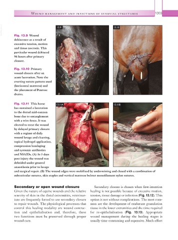
Fig. 13.9 Wound
dehiscence as a result of
excessive tension, motion
and tissue necrosis. This
particular wound dehisced
96 hours after primary
closure.
Fig. 13.10 Primary
wound closure after an
acute laceration. Note the
everting suture pattern used
(horizontal mattress) and
the placement of Penrose
drains.
Fig. 13.11 This horse 13.11A 13.11B
has sustained a laceration
to the dorsal mid-cannon
bone due to entanglement
with a wire fence. It was
elected to treat the wound
by delayed primary closure
with a regime of daily
wound lavage and cleaning,
topical hydrogel application,
compression bandaging
and systemic antibiotics
and NSAIDs. (A) At 5 days
post injury the wound was
debrided under general
anaesthesia prior to lavage
and surgical repair. (B) The wound edges were mobilised by undermining and closed with a combination of
subcuticular sutures, skin staples and vertical mattress bolster monofilament nylon sutures.
Secondary or open wound closure Secondary closure is chosen when first-intention
Given the nature of equine wounds and the relative healing is not possible because of excessive motion,
scarcity of skin in the distal extremities, veterinar- tension, tissue damage or infection (Fig. 13.12). This
ians are frequently forced to use secondary closure option is not without complications. The most com-
to repair wounds. The physiological processes that mon are the development of exuberant granulation
control this healing modality are wound contrac- tissue in the lower extremities and the time required
tion and epithelialisation and, therefore, these for re-epithelialisation (Fig. 13.13). Appropriate
two functions must be preserved through proper wound management during the healing stages is
wound care. usually time-consuming and expensive. Much effort