
Page 1330 - Equine Clinical Medicine, Surgery and Reproduction, 2nd Edition
P. 1330
Wound management and infections of synovial structures 1305
VetBooks.ir 13.14A 13.14B 13.14C
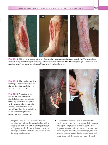
Fig. 13.14 This horse sustained a wound of the medial cannon region 8 days previously (A). The wound was
cleaned, lavaged and bandaged every day, and systemic antibiotics and NSAIDs were given (B). The wound was
repaired by delayed secondary closure (C) and healed without incident.
13.15 13.16
Fig. 13.15 The ‘mesh-expansion’
technique. Note the direction of
the stab incisions parallel to the
direction of the wound.
Fig. 13.16 Protection of the
wound bed with additional
sterile hydrosoluble gel prior to
scrubbing the wound periphery
with a suitable solution, thereby
avoiding contamination of the
wound bed. Note the initial clipping
of the surrounding hair coat.
(Photo courtesy GA Munroe)
• Prepare 1 litre of 0.1% povidone–iodine • Explore the wound in a sterile manner with a
solution and irrigate the wound either with a sterile metal probe or sterile gloved digit to assess
Waterpick™ or with a 60 ml syringe through the depth and presence of wound tracts. It is
a 19-gauge needle. Pressure should be used to important to determine the anatomical structures
dislodge contamination and this can be helped involved, tissue deficits, vascular supply, the level
by using sterile gauzes. of deep contamination, dead space and potential
deep tracts that the wound may have followed.