
Page 1329 - Equine Clinical Medicine, Surgery and Reproduction, 2nd Edition
P. 1329
1304 CHAPTER 13
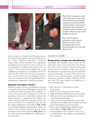
VetBooks.ir 13.12 13.13 Fig. 13.12 An extensive wound
to the dorsal aspect of the tarsal
and proximal metatarsal regions,
typically encountered in barbed-
wire fence injuries. Given its size
and location, this wound is not
amenable to primary closure, and
secondary closure or open wound
healing was selected.
Fig. 13.13 Exuberant
granulation tissue is present
following a wound on the
dorsolateral aspect of the
metatarsal region of a 9-year-old
Quarter horse.
has been made to develop wound-dressing materi- WOUND CLOSURE
als that will significantly shorten the healing period,
but without significant side-effects. Secondary Preparation, lavage and debridement
closure relies on the formation of an appropriate Assessment and treatment of any wound are best
granulation tissue bed for the wound to contract and achieved in a quiet, clean and well-lit environment.
re- epithelialise. Granulation tissue starts to form as The whole horse should be examined thoroughly.
early as 72 hours and proceeds rapidly if the wound Sedation may be necessary in some cases. Prior to
conditions are adequate (i.e. immobility and lack of the administration of any drugs the horse should be
infection). The role of the clinician is to manage an assessed, particularly for signs of shock and severe
open wound in such a way that facilitates the pro- blood loss (e.g. tachycardia, pale or muddy mucous
cesses of wound contraction and epithelialisation by membranes, prolonged capillary refill time [CRT],
maintaining an appropriate wound environment. skin tent). The steps towards the evaluation of a
wound should include:
Delayed secondary closure
Delayed secondary closure is performed by suturing • Move the horse, if necessary, to a better
a wound after the process of fibroplasia has produced environment.
a healthy granulation tissue bed. When applied cor- • Make an initial assessment of the wound.
rectly it can prevent the development of exuberant • Apply sterile hydrosoluble gel to the wound surface.
granulation tissue and considerably accelerate the • Clip hair from the surrounding area to at least
horse’s recovery. In preparation for delayed sec- 5 cm from the wound edges.
ondary wound healing the granulation tissue bed • Lavage the wound with sterile saline, or hose with
must be debrided of its superficial layer and the cold water if not available, while manually removing
skin edges undermined and freshened (Fig. 13.14) gross contamination (e.g. dirt, straw, hair).
to accomplish apposition with minimal tension and • Protect the wound bed with additional sterile
facilitate tissue healing. Additional tension-release hydrosoluble gel (Fig. 13.16) and scrub the
techniques, such as mesh expansion (Fig. 13.15), can wound periphery with a suitable solution, such
be used to decrease the likelihood of dehiscence of as chlorhexidine soap, avoiding contamination of
the suture line. the wound bed.