
Page 1324 - Equine Clinical Medicine, Surgery and Reproduction, 2nd Edition
P. 1324
Wound management and infections of synovial structures 1299
VetBooks.ir for additional features of haemostasis such as the Repair
The repair phase is characterised by the presence
fibrin mesh network and the chemoattraction of
neutrophils. The haemostatic phase may last a few
tive tissue matrix (granulation tissue), and lead to
minutes to a few hours. of fibroblasts, which produce collagen and connec-
wound contraction. The granulation tissue scaffold
Inflammation facilitates the migration of epithelial cells. During
The inflammatory response is characterised mostly second-intention or open wound healing, epitheliali-
by the presence of neutrophils, which are bacteri- sation and contraction are ultimately responsible for
cidal and protect the body against invasion by for- wound repair (Fig. 13.5). This is only possible if the
eign organisms or substances. The release of local wound is clear of debris, infection, tissue necrosis
chemotactic factors serves as a migration signal for and exuberant granulation tissue. Epithelialisation
the neutrophils, which begin a process of diapede- can begin as soon as 12 hours post wounding in pri-
sis and migration to the injury site. Other factors mary healing or after 4 or 5 days in secondary wound
released at a local level (e.g. serotonin, histamine, healing. Wound contraction is most important in
bradykinin) are responsible for a vasodilatory secondary wound closure and stops with contact
response and an increase in vascular permeability, inhibition between cells, excessive tension, exuber-
which further facilitates neutrophil migration. The ant granulation tissue or if full-thickness grafts are
acute inflammatory phase starts within minutes of applied before the 5th day of healing.
a wound occurring and lasts for a few hours to days
depending on the severity of the injury and degree of
contamination. The activation and death of neutro- 13.5
phils is responsible for the release of enzymatic prod-
ucts and inflammatory mediators. In the absence of
infection, neutrophils are not necessary for wound
healing. It is important to emphasise that the appro-
priate balance between acute and chronic inflam-
mation is necessary for a wound to heal optimally
by second intention. While acute inflammation has
been shown to have beneficial effects in wound heal-
ing and should be encouraged, chronic inflammation
promotes the production of exuberant granulation
tissue and should be discouraged.
Debridement
The debridement phase begins within a few hours
of the injury and its duration depends on the sever-
ity of the injury. It starts with the enzymatic break-
down of debris by substances released from the dead
neutrophils. Monocytes are chemoattracted into
the wound at the same time as neutrophils and are
responsible for the elimination of debris, necrotic
tissue, foreign substances and microorganisms.
Unlike neutrophils, monocytes are necessary for
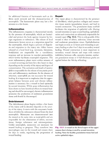
wound healing to progress. They transform into Fig. 13.5 A wound on the dorsal aspect of the upper
macrophages once in the wound and remain there cannon that is being treated by second-intention
for days to weeks, regulating the progression of healing. Note the healthy granulation-tissue bed and
wound healing. active epithelial edges.