
Page 1331 - Equine Clinical Medicine, Surgery and Reproduction, 2nd Edition
P. 1331
1306 CHAPTER 13
VetBooks.ir therapeutic plan should be developed and a decision 13.17
Once wound evaluation has been completed, a
about primary or secondary closure reached. Surgical
debridement is essential and should be performed by
careful resection of necrotic tissue while preserving
all vital structures. Overdebridement may poten-
tially delay the healing process or invade and damage
critical structures such as synovial cavities. Tissues
with questionable viability should be preserved as
much as possible. Occasionally, a skin flap may be
maintained for a few days as a ‘biological’ dressing,
only to be debrided once fibroplasia has started. It is
important to understand that wound debridement is Fig. 13.17 Application of medicinal maggots to a
also a staged procedure that may continue for sev- chronically infected foot abscess.
eral days following initial assessment of the wound.
Any interaction with the wound should be carried 13.18
out with at least clean, if not sterile, gloved hands,
because cross-contamination from the veterinarian’s
hands is possible.
Debridement can also be accomplished by medic-
inal maggot therapy. This involves the use of mag-
gots (larvae of Lucilia sericata) and is a safe, effective
and controlled method of healing chronic wounds
by debridement and disinfection. The maggots are
sterile or disinfected and have a preference to feed on
non-vitalised tissue, purulent exudate and metabolic
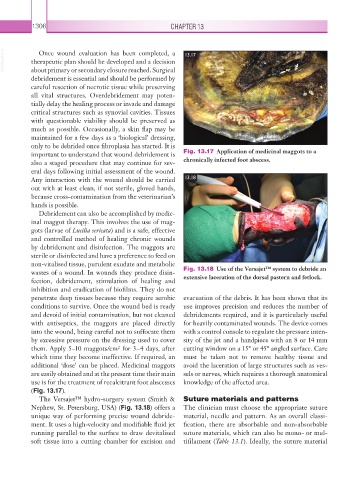
wastes of a wound. In wounds they produce disin- Fig. 13.18 Use of the Versajet™ system to debride an
fection, debridement, stimulation of healing and extensive laceration of the dorsal pastern and fetlock.
inhibition and eradication of biofilms. They do not
penetrate deep tissues because they require aerobic evacuation of the debris. It has been shown that its
conditions to survive. Once the wound bed is ready use improves precision and reduces the number of
and devoid of initial contamination, but not cleaned debridements required, and it is particularly useful
with antiseptics, the maggots are placed directly for heavily contaminated wounds. The device comes
into the wound, being careful not to suffocate them with a control console to regulate the pressure inten-
by excessive pressure on the dressing used to cover sity of the jet and a handpiece with an 8 or 14 mm
them. Apply 5–10 maggots/cm for 3–4 days, after cutting window on a 15° or 45° angled surface. Care
2
which time they become ineffective. If required, an must be taken not to remove healthy tissue and
additional ‘dose’ can be placed. Medicinal maggots avoid the laceration of large structures such as ves-
are easily obtained and at the present time their main sels or nerves, which requires a thorough anatomical
use is for the treatment of recalcitrant foot abscesses knowledge of the affected area.
(Fig. 13.17).
The Versajet™ hydro-surgery system (Smith & Suture materials and patterns
Nephew, St. Petersburg, USA) (Fig. 13.18) offers a The clinician must choose the appropriate suture
unique way of performing precise wound debride- material, needle and pattern. As an overall classi-
ment. It uses a high-velocity and modifiable fluid jet fication, there are absorbable and non-absorbable
running parallel to the surface to draw devitalised suture materials, which can also be mono- or mul-
soft tissue into a cutting chamber for excision and tifilament (Table 13.1). Ideally, the suture material