
Page 1332 - Equine Clinical Medicine, Surgery and Reproduction, 2nd Edition
P. 1332
Wound management and infections of synovial structures 1307
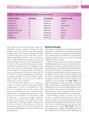
VetBooks.ir Table 13.1 Types of suture material available to the equine clinician
SUTURE MATERIAL ABSORBABLE CONFORMATION COMMERCIAL NAME
Polydioxanone Yes Monofilament PDS/Biosin
Polypropylene No Monofilament Prolene
Polyglactin 910 Yes Multifilament Vicryl/Polysorb
Polyglycolic acid Yes Multifilament Dexon
Polymerised caprolactam No Multifilament Supramid
Poliglecaprone 25 Yes Monofilament Monocryl
Polyester No Multifilament Ethibond
Polyglyconate Yes Monofilament Maxon
used should be user-friendly, prevent a large for- Wound drainage
eign- body reaction, maintain tension and knot The purpose of drainage is to remove the excess fluid
security until the sutured tissue has acquired generated in a wound and to minimise anatomical
enough tension of its own and minimise the risks dead space, thereby decreasing bacterial prolifera-
of harbouring bacteria. This means that a mono- tion, reducing the risk of infection and promoting
filament absorbable material, such as polydioxa- healing. In general, there are two types of draining
none, would be ideal in subcutaneous tissues, and methods: passive and active.
a non-absorbable material, such as polypropylene, Passive drainage mainly uses gravity to facilitate
in the skin. The size of the needle and suture mate- fluid elimination and this can be achieved by strate-
rial should be proportional to the thickness of the gically placed incisions, ventrally located relative to
tissue and, except in subcutaneous tissues, where the drainage area. Gentle body motion helps passive
a round tipped non-cutting needle is preferable, a drainage and should be encouraged whenever possi-
reverse cutting needle is indicated. Suture size 2/0 ble without jeopardising the wound-healing process.
or 3/0 (3 or 3.5 metric) is appropriate for subcutane- In addition, devices such as Penrose drains, made
ous tissues and skin. If excessive tension is antici- out of latex or sterile tubing and not exiting directly
pated, a larger suture size or the use of a different through the wound, may be used to facilitate and
suture pattern may be required. maintain an open drainage flow (Fig. 13.19). The
For wounds where tension is not a concern, a main disadvantage of passive drainage is the possi-
simple or cruciate mattress interrupted pattern bility of promoting an ascending infection. Passive
is appropriate. If tension is anticipated, it is bet- drainage devices are therefore only placed for a short
ter to use a pattern that provides tension relief period of time and in situations where they are abso-
such as a near–far–far–near or a vertical mattress. lutely necessary. The use of passive drains in cases of
Appositional or everting suture patterns are ade- synovial wounds or osteosynthesis is controversial.
quate in the skin. Where the end cosmetic result Active drainage requires a suction system that
is important, a subcuticular simple continuous provides a continuous negative pressure that will
pattern can be chosen. The clinician must bear remove exudate as soon as it is produced and prevent
in mind that the holding strength of this pattern retrograde contamination of the drained area. Active
is inferior to that of a dermal pattern. Closure of drains can be manufactured using a large syringe,
dead space by suturing the subcutaneous layers will some tubing and a needle (Fig. 13.20). They are
prevent the development of seromas and speed the also commercially available. Depending on the exu-
healing process. date volume that is produced, careful emptying and