
Page 692 - Equine Clinical Medicine, Surgery and Reproduction, 2nd Edition
P. 692
Respir atory system: 3.2 Surgical conditions of the respir atory tr act 667
VetBooks.ir 3.121 3.122
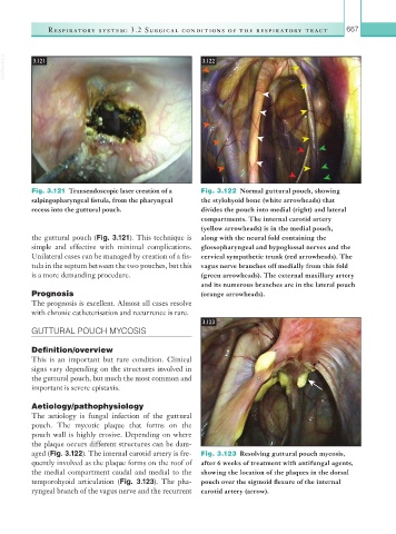
Fig. 3.121 Transendoscopic laser creation of a Fig. 3.122 Normal guttural pouch, showing
salpingopharyngeal fistula, from the pharyngeal the stylohyoid bone (white arrowheads) that
recess into the guttural pouch. divides the pouch into medial (right) and lateral
compartments. The internal carotid artery
(yellow arrowheads) is in the medial pouch,
the guttural pouch (Fig. 3.121). This technique is along with the neural fold containing the
simple and effective with minimal complications. glossopharyngeal and hypoglossal nerves and the
Unilateral cases can be managed by creation of a fis- cervical sympathetic trunk (red arrowheads). The
tula in the septum between the two pouches, but this vagus nerve branches off medially from this fold
is a more demanding procedure. (green arrowheads). The external maxillary artery
and its numerous branches are in the lateral pouch
Prognosis (orange arrowheads).
The prognosis is excellent. Almost all cases resolve
with chronic catheterisation and recurrence is rare.
3.123
GUTTURAL POUCH MYCOSIS
Definition/overview
This is an important but rare condition. Clinical
signs vary depending on the structures involved in
the guttural pouch, but much the most common and
important is severe epistaxis.
Aetiology/pathophysiology
The aetiology is fungal infection of the guttural
pouch. The mycotic plaque that forms on the
pouch wall is highly erosive. Depending on where
the plaque occurs different structures can be dam-
aged (Fig. 3.122). The internal carotid artery is fre- Fig. 3.123 Resolving guttural pouch mycosis,
quently involved as the plaque forms on the roof of after 6 weeks of treatment with antifungal agents,
the medial compartment caudal and medial to the showing the location of the plaques in the dorsal
temporohyoid articulation (Fig. 3.123). The pha- pouch over the sigmoid flexure of the internal
ryngeal branch of the vagus nerve and the recurrent carotid artery (arrow).