
Page 691 - Equine Clinical Medicine, Surgery and Reproduction, 2nd Edition
P. 691
666 CHAPTER 3
VetBooks.ir Aetiology/pathophysiology Differential diagnosis
The aetiology is unclear but probably a congenital
The clinical presentation is quite characteristic.
defect. The ostia are usually patent but are not func-
tioning correctly, allowing a one-way accumulation The most likely confusing differential diagnosis is
a retropharyngeal abscess. These can present with
of air in the pouch. identical pharyngeal swelling, but palpation will
reveal a firm painful mass.
Clinical presentation
Foals usually present with marked swelling of the Diagnosis
parotid region. Palpation reveals a tympanic swell- Radiography is the most valuable technique to con-
ing that is quite painless (Fig. 3.118). Dysphagia or firm the diagnosis (Fig. 3.119). A lateral radiograph
dyspnoea may be present if the swelling has been will reveal the extreme distension of the guttural
allowed to become extreme. pouch. Endoscopy on initial examination will reveal
marked dorsal pharyngeal swelling (Fig. 3.120).
Entry to the guttural pouch causes collapse of the
3.118
pouch and reveals no abnormalities of the internal
structures.
Management
The majority of cases can be managed very suc-
cessfully by chronic catheterisation of the guttural
pouch. A large bore (28Ch) Foley catheter is placed
within the affected guttural pouch(es) and left in
situ for 2–6 weeks. This usually results in sufficient
scarring and alteration of the guttural pouch ostia to
prevent it forming a one-way seal in the future.
A salpingopharyngeal fistula can be created,
using a transendocopic laser to dissect the pha-
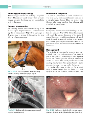
Fig. 3.118 A foal with guttural pouch tympany, ryngeal recess and establish communication into
showing swelling in the pharyngeal region.
3.119 3.120
Fig. 3.119 Radiograph showing a gas-distended Fig. 3.120 Endoscopy of a foal with guttural pouch
guttural pouch (arrows). tympany showing marked dorsal pharyngeal swelling.