
Page 1119 - Adams and Stashak's Lameness in Horses, 7th Edition
P. 1119
Lameness in the Young Horse 1085
The degree of lameness varies from mild and percep
tible only on turns to non‐weight‐bearing. Gross insta
VetBooks.ir fetlock only occurs with biaxial fractures of the body
bility of the suspensory apparatus resulting in a dropped
with extreme distraction. Foals are at the highest risk
for biaxial fractures when turned out to a paddock after
being confined for a period of time. Depending on the
40
fracture configuration, there may be a soft tissue profile
over the involved sesamoid. There is often joint disten
sion, especially with the basilar sesamoid fractures.
Fetlock flexion and direct manual pressure applied over
11
the involved sesamoid usually elicits pain. The diag
nosis is confirmed by radiographs and may be facili
tated by ultrasound evaluation.
Treatment for proximal sesamoid bone fractures in
foals is conservative and consists of confinement to a
stall‐sized area for 4–8 weeks, followed by a graduated
turnout schedule. The prognosis for future athleticism is
good, but the sesamoid commonly heals in an elongated
manner, which is often criticized if the horse is sold at
public auction.
Only in rare instances is external coaptation with a
cast or splint indicated. Most devices are contraindicated
40
for fear of promoting laxity of the flexor tendons.
Likewise, surgical repair is usually futile and applies to
salvage cases with total compromise of the suspensory
apparatus that have marked distraction of the fracture
fragments and dropping of the fetlock. The logical
surgical technique is circumferential wiring or suturing
of the fractured sesamoid bones. External coaptation
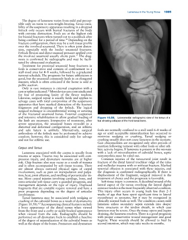
and intensive rehabilitation to allow gradual loading of Figure 10.58. Lateromedial radiographic view of the tarsus of a
the limb are necessary. Irrespective of treatment, after foal showing collapse of the third tarsal bone.
severe separation, the sesamoid bones remodel in a
distorted and deformed configuration, and an athletic
and sale future is unlikely. Alternatively, surgical foals are normally confined to a stall until 6–8 weeks of
arthrodesis of the fetlock may be performed to achieve age or until acceptable mineralization has occurred to
comfort; however, this is considered only for salvage minimize wedging or crushing. Tarsal wedging or
and not for athletic use. crushing usually does not cause lameness in the neonate.
Gait abnormalities are recognized only after periods of
exertion following turnout with other foals or after ath
Carpus and Tarsus letic activity begins. If lameness is present in the neonate
Lameness associated with the carpus is usually from with a lack of mineralization of cuboidal bones, septic
trauma or sepsis. Trauma may be associated with com osteomyelitis must be ruled out.
pression injury, and dysmature neonates are at higher Common injuries of the tarsocrural joint result in
risk. Chip fracture also may occur as a result of trauma fracture of the distal lateral trochlear ridge of the talus
and is often accompanied by soft tissue trauma. There and malleolar trauma with or without fracture. Marked
are almost always outward physical signs of carpal synovial effusion is associated with these injuries, and
involvement, such as pain on manipulation and palpa the diagnosis is confirmed radiographically. If there is
tion, heat, joint effusion, and swelling of periarticular tis displacement of the fragment, surgical removal is the
sue. Most carpal injuries involving cartilage, bone, and treatment of choice and the prognosis is favorable.
intra‐articular soft tissue carry a guarded prognosis, and Soft tissue injury is common. A decubital wound of the
management depends on the type of injury. Displaced lateral aspect of the tarsus overlying the lateral digital
fragments that are unstable require removal and have a extensor tendon is the most frequently observed condition.
poor prognosis depending on the degree of articular This injury often occurs in young foals less than 2–3
involvement. weeks of age that have spent significant time in lateral
Tarsal lameness may be associated with wedging or recumbency as neonates, but it may be recognized in
crushing of the cuboidal bones as a result of dysmaturity clinically normal foals as well. The condition causes mild
8,9
(Figure 10.58). Accompanying clinical features include lameness unless secondary sepsis extends into deeper
a boxy appearance of the distal tarsus when viewed bony structures or the tendon sheath, in which case
from the front and a curby or sickle‐hocked appearance lameness may be severe. Once the tissue opens and begins
when viewed from the side. Radiographs should be draining, the lameness resolves. There is a good prognosis
performed on all dysmature foals to establish a baseline with proper conservative wound management and good
of the degree of mineralization of the cuboidal bones as hygiene. These wounds should be allowed to heal by
well as the shape of the bones. Premature and dysmature second intention, which may take weeks to months.