
Page 1116 - Adams and Stashak's Lameness in Horses, 7th Edition
P. 1116
1082 Chapter 10
digital compression and flexion or manipulation of the Foot Lameness
joint. The surfaces of the proximal sesamoid bones dur Distal phalangeal fractures in horses have been clas
VetBooks.ir areas. Sequential and methodical palpation of the entire sified into seven types. 10,40 Although any of these may
ing extension and flexion are palpated for sensitive
occur in foals, type VII fractures are unique to foals and
limb including surfaces of the articular as well as non
articular structures should be routinely performed. occur very commonly on Thoroughbred breeding oper
Most articular structures affected with sepsis yield a ations. There is a reported prevalence of 19%–74%
response of the foal with digital compression of the that are recognized predominantly between 2 weeks
5,13,48
joint margins and should raise suspicions of the clini and 5 months of age. Fractures involve the nonar
cian. Likewise, focal areas of injury typically produce a ticular margin dorsal to the palmar/plantar process.
pain response to digital compression and warrant fur They have been described as ossicles or osseous bodies
25
ther investigation with imaging procedures. Deep pal but are commonly referred to as “wing” fractures. The
pation of the proximal forelimb, vertebral column, lateral palmar process is almost always involved, but
proximal hindlimbs, and pelvis should be a routine part occasionally they occur biaxial or bilateral; seldom is
of the examination. Each compartment of the stifle only the medial wing involved. Although factors such as
should be isolated and palpated for effusion, pain, and hard or soft surfaces, changes in turnout schedules, and
temperature alteration. The entire rib cage should be hoof conformation and application of external hoof
carefully examined especially in the young foal that acrylics are incriminated, no single factor has been sub
13,14
may have sustained rib fracture with subsequent fore stantiated as causative. Because many type VII frac
limb lameness. tures are recognized as incidental findings, they have
Isolation of the lame limb and the specific origin of been considered separate centers of ossification; how
40
pain may be challenging if subtle, but in general foals ever this has not been documented.
readily display locomotor evidence of the location of The degree of lameness associated with type VII frac
pain. Most foals attempt to avoid the painful compo tures varies from non‐apparent to non‐weight‐bearing
nent of the gait and usually display more exaggerated but generally display a 3/5 lameness acutely that
lameness reactions than adults. Observation of the improves in a matter of days. While ambulating in a
young foal at rest and unrestrained in the company of straight line, the foal will display a characteristic abduc
the mare may be useful in detecting mild gait altera tion of the limb during contact with the ground, pre
tions or postural changes. Special attention should be sumably to reduce loading on the painful region. The
given to the stance of the foal, particularly any trends lameness is more exaggerated on turns as well.
in abnormal posture. Young, untrained foals may There is usually a palpable increase in the intensity
require gait evaluation while unrestrained and follow of the digital pulse, but only a mild increase in the tem
ing the mare. The gait should be assessed at a slow perature of the foot. The most consistent clinical find
walk in a straight line and in turns in both directions. ing is an increase in reaction to medial‐to‐lateral
Placement of the foot as well as limb and body carriage compression of the hoof wall at the quarters and heels
during ambulation is important in localizing lameness. (Figure 10.54). Palmar digital anesthesia of the involved
Although there are some common findings with certain side will eliminate the lameness. The diagnosis is con
conditions (see below), there are few absolutes regard firmed radiographically using the 45° dorsal–ventral
ing gait characteristics and location of lameness in
foals. Conventional lameness diagnostic anesthesia is
often necessary to isolate the location of the source of
lameness.
NONINFECTIOUS CAUSES OF LAMENESS
There are a multitude of events that may affect any
part of the musculoskeletal system resulting in lame
ness; several disorders are unique to foals and occur
routinely. Categorically lameness may be divided into
infectious or noninfectious causes. Noninfectious causes
include trauma, metabolic (DOD), or afflictions of
vascular or neurogenic origin. For discussion purposes
disorders causing lameness will be mentioned by ana
tomical region involved.
Virtually all components of the musculoskeletal sys
tem are susceptible to injury and lameness. Intrinsic and
external sources of trauma commonly result in struc
tural compromise to tissue of the musculoskeletal sys
tem. Other conditions resulting in lameness include
those associated with DOD and disorders of vascular or
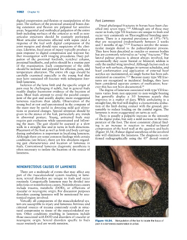
neurogenic origin. Several disorders specific to foals Figure 10.54. Manipulation of the foot to locate the focus of
occur routinely and are worth mention. pain in a lameness examination in a foal.