
Page 1113 - Adams and Stashak's Lameness in Horses, 7th Edition
P. 1113
Lameness in the Young Horse 1079
VetBooks.ir
B
A
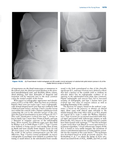
Figure 10.53. (A) Flexed lateral–medial radiograph and (B) caudal–cranial radiograph of subchondral cystic lesions (arrows in A) of the
medial femoral condyle of the femur.
of importance are the distal metacarpus or metatarsus in noted in the limb contralateral to that of the clinically
the fetlock joint, the distal proximal phalanx in the prox significant SCL, and type 4 lesions were defined as those
imal interphalangeal joint, and shoulder, elbow, carpus, that had a lucency in the condyle with or without an
distal phalanx, and talus. Principles of diagnosis and articular defect but no radiographic evidence of an
treatment will be restricted here to the most common opening in the subchondral bone plate (less common).
entity, the medial condyle of the femur. Digital radiographs and surveys have led to increased
With regard to radiographic appearance and classifi scrutiny of radiographs, and type 1 lesions have now
cation of SCLs of the MFC, there has been an evolution. evolved into two sizes of concave defects as well as
Initially there were two types: type 1 was a radiographi including flattening of the condyle.
cally domed‐shaped lucent area that was confluent with The decision for surgery based on the authors’ expe
a flattened joint surface, and type 2 had a circular lucent rience is based on the presence or absence of clinical
area within the condyle with a thinner radiolucent track signs. Most cases with clinical signs referable to the
connecting the cyst to the articular surface of the con medial femoral tibia joint when there is an SCL present
29
dyle (the typical manifestation as seen in Figure 10.53). are typical SCLs (type 2 under the Wallace classifica
This early classification evolved into type 1, 10 mm or tion). Type 1 lesions are occasional associated with clini
less in depth, type 2 more than 10 mm in depth, and type cal signs and treated with arthroscopic surgery as well.
3 flattened or irregular contours of the subchondral Historically, SCL lesions in the medial femoral condyle
bone. More recently five different types were described have been treated with curettage, 5,24,29 but this technique
by Wallace et al. in 2008 (Figure 5.127). Type 1 lesions is quite rarely done now. Based on recent agreement to a
27
were defined as being concave and smaller than 10 mm treatment algorithm, it had arthroscopic surgery. If the
14
in depth and were usually dome shaped. Types 2A and SCL is found to have stable margins, the treatment of
2B were typical cystic lesions over 10 mm in depth, and choice is intralesional injection of triamcinolone aceton
the width of the surface communication was the only ide into the contents of the cystic lesion. This technique
27
difference. Type 3 lesions (noted incidentally on survey was developed based on work previously cited by von
radiographs of yearlings) were defined as condylar flat Rechenberg et al., in which the lining of SCLs pro
20
tening or small defects in the subchondral bone usually duced increased levels of PGE , MMPs, and NO, as well
2