
Page 1109 - Adams and Stashak's Lameness in Horses, 7th Edition
P. 1109
Lameness in the Young Horse 1075
VetBooks.ir
A B
C D
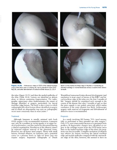
Figure 10.49. Arthroscopic views of OCD of the lateral trochlear lesion on the medial trochlear ridge of the femur manifesting as
ridge of the femur prior to probing (A), during elevation of the OCD elevated cartilage of normal thickness without a subchondral defect
flap (B), and after debridement of osteochondritic tissue (C). (D) A beneath.
the talus (Figure 10.51) and then the medial malleolus of Warmblood tarsocrural joints showed development (and
the tibia (Table 10.4). Lesions are identified as defects resolution in most cases) of lesions of the DIRT and lat
alone or defects containing fragmentation. The radio eral trochlear ridge of the talus over the first 5 months of
3
graphic appearance often underestimates the extent of life. Surgery should be considered early enough in the
damage identified at surgery, particularly for lateral course of the disease (but after 5 months of age) so that
trochlear ridge lesions. The hock is also a joint in which the joint capsule is not unduly stretched, which makes
radiographically silent lesions (those identified at surgery resolution of the joint effusion less likely. Arthroscopic
and in which no abnormality was seen on radiographs) surgery with removal of fragments and debridement of
occur more commonly than in other joints. 13 defective tissue is recommended.
Treatment Prognosis
Although lameness is usually minimal with hock In a study involving 183 horses, 76% raced success
13
OCD, surgery is the recommended treatment. Lameness fully or performed at their intended use after surgery.
may only be a problem at racing speeds, or at upper lev Only 11% were unsuccessful because of a persistent hock
els of performance, and this cannot be determined during problem. If degenerative changes were identified at sur
a clinical examination. Resolution of the effusion cannot gery in the cartilage remote from the OCD lesion (wear
be expected without removal of the abnormal tissue. lines on the medial trochlear ridge of the talus), the prog
However, not all horses need surgery. Those with small nosis was less favorable. Complete resolution of effusion
lesions, minimal effusion, no lameness, and a potential was inferior for lesions involving the lateral trochlear
career as a pleasure horse or light use horse may not ridge and medial malleolus compared with the intermedi
require surgery. Sequential radiographs of Dutch ate ridge of the tibia; however, this seemed to have no