
Page 1107 - Adams and Stashak's Lameness in Horses, 7th Edition
P. 1107
Lameness in the Young Horse 1073
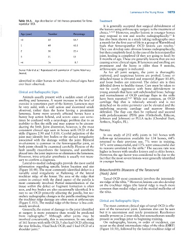
Table 10.3. Age distribution of 161 horses presented for femo- Treatment
ropatellar OCD. It is generally accepted that surgical debridement of
VetBooks.ir Age (year) Number Percentage the lesions using arthroscopic surgery is the treatment of
However, smaller lesions in younger horses
choice.
4,11,15
15
may respond to rest and resolve radiographically. It
<1 22 13.7 has also been shown in a study taking radiographs once
a month for the first year of life in a group of Warmblood
3
1 68 42.2 foals that femoropatellar OCD lesions can resolve.
They can develop into obvious lesions radiographically,
2 36 22.4 but then completely heal. In the case of the femoropatellar
joint, healing is completed (if they are going to heal) by
3 21 13
8 months of age. These are generally lesions that are not
>4 14 8.7 causing severe clinical signs. If lameness and swelling are
prominent and the horse is older than 8 months,
arthroscopic surgery is indicated.
Source: Foland et al. Reproduced with permission of Equine Veterinary
4
Journal. As for all joint surgery, the joint is thoroughly
explored, and suspicious lesions are probed. Loose or
detached tissue is elevated and removed (Figure 10.49),
identified in older horses in which no clinical signs have and loose bodies are removed. The defect site is then
ever been observed. debrided down to healthy tissue. Care must be taken to
not be overly aggressive with bone debridement in
Clinical and Radiographic Signs young animals that have soft subchondral bone. Salvage
and reattachment of OCD cartilage flaps in the stifle
Animals usually present with a sudden onset of joint 15,17
swelling and lameness. A recent increase in the level of have been reported. While not common, an OCD
cartilage flap that is relatively smooth and is not
exercise is sometimes part of the history. Lameness may detached on its entire perimeter can be elevated and the
be very mild, with a stiff action and shortened stride underlying necrotic cartilage and marrow fibrosis
observed, rather than the horse having a prominent debrided. The flap can then be replaced and secured
lameness. Some more severely affected horses have a with polydioxanone (PDS) pins (OrthoSorb, Ethicon,
bunny hop action behind, and severe cases can some Johnson and Johnson) or PLLA tacks (Chondral Dart,
times be confused with a neurologic problem due to an Arthrex, Naples, FL).
inability to flex the stifle and may appear to be circum
ducting the limb. Joint distention, however, is the most
consistent clinical sign seen in horses with OCD of the Prognosis
stifle (Figures 2.90 and 5.110). Careful palpation of the In one study of 252 stifle joints in 161 horses with
joint may identify free bodies or the surface irregularity follow‐up information available for 134 horses, 64%
associated with the damage within the joint. Bilateral returned to their previous use, 7% were in training,
involvement is common in the femoropatellar joint, so 16% were unsuccessful, and 13% were unsuccessful due
both joints should be examined carefully. Flexion of the to reasons unrelated to the stifle. The success rate was
4
limb usually exacerbates the lameness, and anesthetic higher in horses with smaller lesions and in older horses.
placed into the joint improves or eliminates the lameness. However, the age factor was considered to be due to the
However, intra‐articular anesthesia is usually not neces fact that the most severe lesions were generally identified
sary to confirm a diagnosis. in younger horses.
Lateral‐to‐medial radiographs provide the most useful
information regarding specific lesion location and size
(Figure 10.48). The most commonly identified defect is a Osteochondritis Dissecans of the Tarsocrural
variably sized irregularity or flattening of the lateral (Hock) Joint
trochlear ridge of the femur. The area of the ridge that
comes in contact with the distal aspect of the patella is Tarsal OCD most commonly involves the intermedi
13
most commonly involved. Partial mineralization of the ate ridge of the tibia. However, lesions can also develop
tissue within the defect or fragment formation is often on the trochlear ridges (the lateral ridge is much more
seen, and free bodies are also occasionally identified. It is common than medial ridge) and the medial malleolus of
rare to see OCD primarily affecting the patella, but sec the tibia.
ondary radiographic changes in the patella resulting from
the trochlear ridge damage are often seen at arthroscopy Clinical and Radiographic Signs
(Figure 5.113). The medial ridge of the femur is less com
monly involved. The most common clinical sign of tarsal OCD is effu
Generally, the extent of damage to the joint identified sion of the tarsocrural joint. Lameness also can be seen
at surgery is more extensive than would be predicted but it is uncommon and rarely prominent. Racehorses
from radiographs. Although other joints may be usually present as 2‐year‐olds, but nonracehorses usually
25
involved concurrently, this is uncommon. In one study present as yearlings prior to beginning training.
of 161 horses with stifle OCD, 5 also had OCD affecting On radiographs, lesions, in order of incidence, can
the rear fetlocks, 4 had hock OCD, and 1 had OCD of a occur on the distal intermediate ridge of the tibia (DIRT)
shoulder joint. 4 (Figure 10.50), followed by the lateral trochlear ridge of