
Page 1112 - Adams and Stashak's Lameness in Horses, 7th Edition
P. 1112
1078 Chapter 10
VetBooks.ir
A B
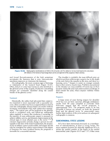
Figure 10.52. Radiographic manifestations of OCD of the shoulder. (A) This defect in the humeral head is the only lesion.
(B) Defects in the humeral head (large black arrow) and glenoid of the scapula (2 black arrows).
and forced flexion/extension of the limb sometimes The shoulder is probably the most difficult joint on
accentuates the lameness that is seen. Intra‐articular which to perform arthroscopic surgery, due to the depth
anesthesia improves or eliminates the lameness. of the joint below the muscles in the area, and there is a
On radiographs, the most common sign is flattening learning curve. Surgery is easier on younger animals
15
or indentation of the humeral head (Figure 10.52). due to their relative muscle mass. Problems encountered
Often, defects or cystic‐type lesions are also identified in in the shoulder are inaccessibility of lesions due to their
the glenoid cavity of the scapula. Productive remodeling location within the joint and extravasation or leakage of
changes are commonly identified along the caudal fluid outside the joint, which impairs visibility within
border of the glenoid cavity. 2 the joint.
Prognosis
Treatment
A large series of cases having surgery for shoulder
Historically, the author had advocated that conserva OCD has not yet been reported, although preliminary
tive treatment is never associated with a successful out results from 80 cases compiled at Colorado State
come. More recently it has been recognized that in selected University suggest that the overall prognosis is
cases the radiographic obvious lesion can resolve without approximately 50%. The prognosis seems to be less
secondary osteoarthritis (OA) with conservative manage favorable if lesions are present on both the humeral
ment. The typical case will be localized to the glenoid, head and the glenoid cavity. In unsuccessful cases,
15
which is recognized early, and follow‐up radiographs further deterioration of the joint surfaces on subsequent
taken regularly to ensure that resolution is occurring. In radiographs is common.
the majority of cases arthroscopic surgery is necessary to
achieve athletic activity, and dramatic improvement in the
clinical signs in most affected cases is the norm. 2,15,16 If SUBCHONDRAL CYSTIC LESIONS
extensive degenerative arthritic change is present on radi
ographs at the time of the initial examination, the progno SCLs have been mentioned previously as a manifesta
sis for an athletic career is unfavorable, and surgery should tion of osteochondrosis, at least in young horses. The
only be considered for relative improvement in the degree most common location for clinically significant SCLs is
of lameness. For more localized lesions, the prognosis is within the medial condyle of the femur in the medial
favorable for a successful outcome. femorotibial joint (Figures 10.53 and 7.17). Other areas