
Page 1117 - Adams and Stashak's Lameness in Horses, 7th Edition
P. 1117
Lameness in the Young Horse 1083
VetBooks.ir
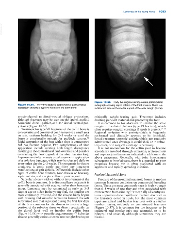
Figure 10.56. Forty five degrees dorsoproximal palmarodistal
Figure 10.55. Forty five degrees dorsoproximal palmarodistal radiograph showing septic osteitis of the third phalanx. There is a
radiograph showing a type VII fracture of the coffin bone. radiolucent area on the medial aspect of the solar margin (arrow).
proximolateral to distal–medial oblique projections, minimally weight‐bearing gait. Treatment includes
although fractures may be seen on the lateral–medial, draining purulent material and protecting the foot.
horizontal dorsal–palmar, and 45° dorsal–ventral pro It is common to for abscesses to involve the solar
jections (Figure 10.55). margin of the distal phalanx (type VI fracture), which
Treatment for type VII fractures of the coffin bone is often requires surgical curettage if sepsis is present. 34–37
conservative and consists of confinement to a small area Regional perfusion with antimicrobials is frequently
on soft, uniform bedding for 2–3 weeks or until the performed and clinically appears to be beneficial.
horse is comfortable enough for paddock turnout. Broad‐spectrum systemic antimicrobials are routinely
48
Rigid coaptation of the foot with a shoe is unnecessary administered once drainage is established, or in refrac
but has become popular. Two complications of shoe tory cases, or if surgical curettage is necessary.
application include creating limb length discrepancy It is not uncommon for the coffin joint to become
resulting in the contralateral limb overload and possibly secondarily involved through extension; arthrocentesis
contracting the hoof capsule if the shoe remains long. and copious joint lavage are indicated in addition to the
Improvement in lameness is usually seen with application above treatments. Generally, with joint involvement
of a soft foot bandage, which may be changed daily or subsequent to hoof abscess, there is a guarded to poor
every other day for 2–3 weeks. The prognosis for future prognosis because this is often associated with an
soundness is good; rarely are there any long‐term aggressive and rapidly spreading infection.
ramifications or gait deficits. Differentials include other
types of coffin bone fracture, foot abscess or bruising,
septic osteitis, and a septic coffin or pastern joint. Proximal Sesamoid Bone
Subsolar abscess with or without involvement of the Fracture of the proximal sesamoid bones is another
coffin bone is common. It is discussed here because it is common lameness condition on commercial breeding
generally associated with trauma rather than hematog farms. These are most commonly seen in foals younger
enous. Lameness may be recognized as early as 3–5 than 6–8 weeks of age; they are often associated with
days of age or older. In the young foal, the hindfeet are overexertion from running. Essentially all configura
11
more commonly involved, and the abscess likely begins tions of proximal sesamoid bone fracture described in
with bruising and subsolar hematoma beneath the soft the literature occur in foals, but the most common
keratinized sole that is present during the first few days types are apical and basilar fractures with a smaller
of life. It is common for the abscess to involve a large number having midbody or comminuted fractures
portion of the subsolar tissue or dissect proximally up (Figure 10.57). It is common for the fractures to be
the dorsal hoof wall or involve the coffin bone unilateral and involve only one sesamoid, or to be
(Figure 10.56) with possible sequestration. 2,37 Subsolar bilateral and uniaxial, although sometimes they are
abscess generally causes a severe non‐weight‐bearing or biaxial.