
Page 1120 - Adams and Stashak's Lameness in Horses, 7th Edition
P. 1120
1086 Chapter 10
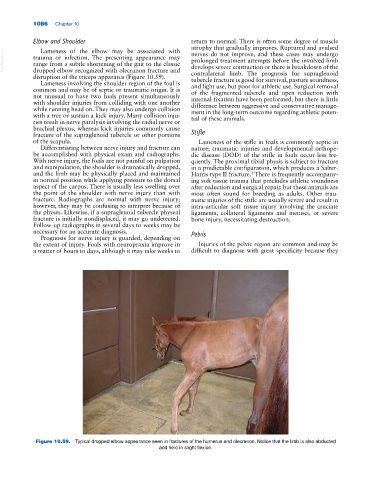
Elbow and Shoulder return to normal. There is often some degree of muscle
atrophy that gradually improves. Ruptured and avulsed
Lameness of the elbow may be associated with
VetBooks.ir trauma or infection. The presenting appearance may prolonged treatment attempts before the involved limb
nerves do not improve, and these cases may undergo
range from a subtle shortening of the gait to the classic
develops severe contraction or there is breakdown of the
dropped elbow recognized with olecranon fracture and
disruption of the triceps apparatus (Figure 10.59). contralateral limb. The prognosis for supraglenoid
tubercle fracture is good for survival, pasture soundness,
Lameness involving the shoulder region of the foal is and light use, but poor for athletic use. Surgical removal
common and may be of septic or traumatic origin. It is of the fragmented tubercle and open reduction with
not unusual to have two foals present simultaneously internal fixation have been performed, but there is little
with shoulder injuries from colliding with one another difference between aggressive and conservative manage
while running head on. They may also undergo collision ment in the long‐term outcome regarding athletic poten
with a tree or sustain a kick injury. Many collision inju tial of these animals.
ries result in nerve paralysis involving the radial nerve or
brachial plexus, whereas kick injuries commonly cause
fracture of the supraglenoid tubercle or other portions Stifle
of the scapula. Lameness of the stifle in foals is commonly septic in
Differentiating between nerve injury and fracture can nature; traumatic injuries and developmental orthope
be accomplished with physical exam and radiographs. dic disease (DOD) of the stifle in foals occur less fre
With nerve injury, the foals are not painful on palpation quently. The proximal tibial physis is subject to fracture
and manipulation, the shoulder is dramatically dropped, in a predictable configuration, which produces a Salter‐
and the limb may be physically placed and maintained Harris type II fracture. There is frequently accompany
3
in normal position while applying pressure to the dorsal ing soft tissue trauma that precludes athletic soundness
aspect of the carpus. There is usually less swelling over after reduction and surgical repair, but these animals are
the point of the shoulder with nerve injury than with most often sound for breeding as adults. Other trau
fracture. Radiographs are normal with nerve injury; matic injuries of the stifle are usually severe and result in
however, they may be confusing to interpret because of intra‐articular soft tissue injury involving the cruciate
the physes. Likewise, if a supraglenoid tubercle physeal ligaments, collateral ligaments and menisci, or severe
fracture is initially nondisplaced, it may go undetected. bone injury, necessitating destruction.
Follow‐up radiographs in several days to weeks may be
necessary for an accurate diagnosis. Pelvis
Prognosis for nerve injury is guarded, depending on
the extent of injury. Foals with neuropraxia improve in Injuries of the pelvic region are common and may be
a matter of hours to days, although it may take weeks to difficult to diagnose with great specificity because they
Figure 10.59. Typical dropped elbow appearance seen in fractures of the humerus and olecranon. Notice that the limb is also abducted
and held in slight flexion.