
Page 1111 - Adams and Stashak's Lameness in Horses, 7th Edition
P. 1111
Lameness in the Young Horse 1077
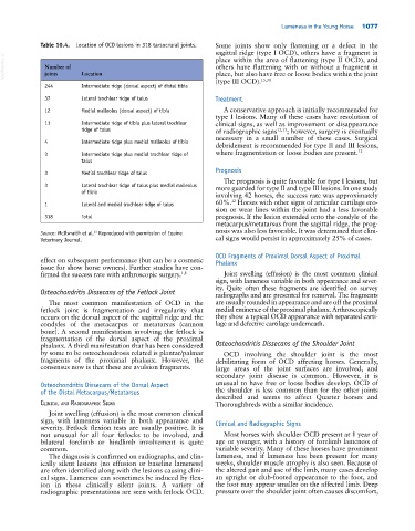
Table 10.4. Location of OCD lesions in 318 tarsocrural joints. Some joints show only flattening or a defect in the
sagittal ridge (type I OCD), others have a fragment in
VetBooks.ir Number of Location others have flattening with or without a fragment in
place within the area of flattening (type II OCD), and
place, but also have free or loose bodies within the joint
joints
244 Intermediate ridge (dorsal aspect) of distal tibia (type III OCD). 15,30
37 Lateral trochlear ridge of talus Treatment
12 Medial malleolus (dorsal aspect) of tibia A conservative approach is initially recommended for
type I lesions. Many of these cases have resolution of
11 Intermediate ridge of tibia plus lateral trochlear clinical signs, as well as improvement or disappearance
ridge of talus of radiographic signs 12,15 ; however, surgery is eventually
necessary in a small number of these cases. Surgical
4 Intermediate ridge plus medial malleolus of tibia
debridement is recommended for type II and III lesions,
3 Intermediate ridge plus medial trochlear ridge of where fragmentation or loose bodies are present. 12
talus
Prognosis
3 Medial trochlear ridge of talus
The prognosis is quite favorable for type I lesions, but
3 Lateral trochlear ridge of talus plus medial malleolus more guarded for type II and type III lesions. In one study
of tibia
involving 42 horses, the success rate was approximately
12
1 Lateral and medial trochlear ridge of talus 60%. Horses with other signs of articular cartilage ero
sion or wear lines within the joint had a less favorable
318 Total prognosis. If the lesion extended onto the condyle of the
metacarpus/metatarsus from the sagittal ridge, the prog
nosis was also less favorable. It was determined that clini
13
Source: McIlwraith et al. Reproduced with permission of Equine
Veterinary Journal. cal signs would persist in approximately 25% of cases.
OCD Fragments of Proximal Dorsal Aspect of Proximal
effect on subsequent performance (but can be a cosmetic Phalanx
issue for show horse owners). Further studies have con
firmed the success rate with arthroscopic surgery. 1,8 Joint swelling (effusion) is the most common clinical
sign, with lameness variable in both appearance and sever
ity. Quite often these fragments are identified on survey
Osteochondritis Dissecans of the Fetlock Joint radiographs and are presented for removal. The fragments
The most common manifestation of OCD in the are usually rounded in appearance and are off the proximal
fetlock joint is fragmentation and irregularity that medial eminence of the proximal phalanx. Arthroscopically
occurs on the dorsal aspect of the sagittal ridge and the they show a typical OCD appearance with separated carti
condyles of the metacarpus or metatarsus (cannon lage and defective cartilage underneath.
bone). A second manifestation involving the fetlock is
fragmentation of the dorsal aspect of the proximal
phalanx. A third manifestation that has been considered Osteochondritis Dissecans of the Shoulder Joint
by some to be osteochondrosis related is plantar/palmar OCD involving the shoulder joint is the most
fragments of the proximal phalanx. However, the debilitating form of OCD affecting horses. Generally,
consensus now is that these are avulsion fragments. large areas of the joint surfaces are involved, and
secondary joint disease is common. However, it is
Osteochondritis Dissecans of the Dorsal Aspect unusual to have free or loose bodies develop. OCD of
of the Distal Metacarpus/Metatarsus the shoulder is less common than for the other joints
described and seems to affect Quarter horses and
CliniCal and radiograPhiC signs Thoroughbreds with a similar incidence.
Joint swelling (effusion) is the most common clinical
sign, with lameness variable in both appearance and Clinical and Radiographic Signs
severity. Fetlock flexion tests are usually positive. It is
not unusual for all four fetlocks to be involved, and Most horses with shoulder OCD present at 1 year of
bilateral forelimb or hindlimb involvement is quite age or younger, with a history of forelimb lameness of
common. variable severity. Many of these horses have prominent
The diagnosis is confirmed on radiographs, and clin lameness, and if lameness has been present for many
ically silent lesions (no effusion or baseline lameness) weeks, shoulder muscle atrophy is also seen. Because of
are often identified along with the lesions causing clini the altered gait and use of the limb, many cases develop
cal signs. Lameness can sometimes be induced by flex an upright or club‐footed appearance to the foot, and
ion in these clinically silent joints. A variety of the foot may appear smaller on the affected limb. Deep
radiographic presentations are seen with fetlock OCD. pressure over the shoulder joint often causes discomfort,