
Page 400 - Adams and Stashak's Lameness in Horses, 7th Edition
P. 400
366 Chapter 3
Harris fractures and subluxations can also occur. These
injuries are usually diagnosed clinically and radiograph
VetBooks.ir or minimally displaced; in such cases scintigraphy may
ically. However, fractures can sometimes be very small
add useful information in the diagnosis. Osteomyelitis
secondary to trauma to the proximal radius with possi
ble progression to septic elbow arthritis has been docu
mented as a large and intense area of radiotracer in that
region. Radiographically, these lesions may be subtle
86
or not apparent if the infection has not advanced enough
to cause substantial bone lysis.
Subchondral bone cysts and cartilage lesions may not
show scintigraphic abnormalities unless the underlying
subchondral bone is affected.
Shoulder and Scapula
Injuries to the shoulder and scapula in the horse are
uncommon. Fractures of the greater tubercle may occur,
but frequently the diagnosis is based on clinical evalua
tion in combination with radiography or ultrasonog
raphy. 57,94 An area of increased radiotracer in the
cranioproximal humerus may represent a fracture of the
tubercle(s) or extension of an infectious/inflammatory
process related to the bicipital bursa (Figure 3.182). An
unusual case of abnormal radiotracer in the cranioprox
imal humerus corresponding to a cyst‐like lesion of the
intermediate tubercle in absence of bicipital bursitis was
reported. In general, subchondral bone cysts and small
67
cartilage defects are lesions that are not detected
scintigraphically unless there are inflammatory changes
extending to the adjacent subchondral bone.
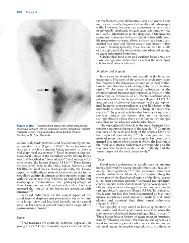
Figure 3.180. Delayed phase lateral view of the left humerus, The supraglenoid tubercle is the most frequent loca
showing a focal and intense radiotracer on the craniodistal humeral tion for a traumatic fracture of the scapula. 19,20 Complete
diaphysis (arrow), consistent with a stress fracture. Source: fractures of the neck and body of the scapula have also
Courtesy of Dr. Ryan Carpenter. been reported sporadically. 19,20 The scintigraphic diag
nosis of stress fractures of the scapula has been docu
caudodistal, caudoproximal, and less commonly cranio mented in a report of two cases in which the location of
proximal cortices (Figure 3.180). Stress fractures of the focal and intense radiotracer corresponding to the
62
the radius are less common being reported to have a fractures was located in the caudal midbody and the
15
mid‐diaphyseal occurrence. Focal increase radiophar ventral aspect of the neck, respectively.
52
maceutical uptake in the medullary cavity of long bones
was first described as “bone infarcts” and subsequently Tarsus
68
as enostosislike lesions (Figure 3.181). 7,66 These lesions
are frequently seen in the tibia, radius, humerus, and Distal tarsal radiotracer is usually seen in jumping
MCIII/metatarsal bones. Radiographically, the lesions horses, followed by racing Standardbreds and less com
5,29,30
appear as well‐defined areas of increased opacity in the monly Thoroughbreds. The increased radiotracer
medullary cavities. It appears to be a transient condition can be unilateral or bilateral; a distribution along the
with the lesions resolving in follow‐up radiographic and entire area of the distal tarsal region of the dorsal aspect
60
scintigraphic examinations. The pathophysiology of is the most common pattern. Increased radiopharma
these lesions is not well understood, and it has been ceutical uptake in the distal tarsal region corresponds to
reported that not all of the lesions are associated with OA or degenerative changes that may or may not be
lameness. 7,65 radiographically apparent (Figure 3.183). Talocalcaneal
Increased radiotracer on the caudal aspect of the OA is rare but has also been documented as focal and
distal femoral diaphysis just proximal to the stifle joint intense radiotracer predominantly medially and more
on a lateral view and localized laterally on the caudal plantar and proximal than distal tarsal radiotracer
77
view has been seen in cases of injury to the origin of the (Figure 3.184).
gastrocnemius muscle. 87 Scintigraphy is very useful in localizing fractures of
the central and third tarsal bones, especially when the
fracture is not displaced, hence radiographically occult.
84
Elbow These horses have a history of acute onset of lameness,
usually following exercise. The fracture will appear as a
Ulnar fractures are relatively common, especially in focal and intense region of radiotracer in the mid to dis
young horses. Other traumatic injuries such as Salter– tal tarsal region. Incomplete sagittal fracture of the talus
14