
Page 405 - Adams and Stashak's Lameness in Horses, 7th Edition
P. 405
Diagnostic Imaging 371
VetBooks.ir
A B
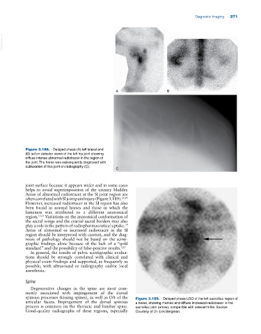
Figure 3.188. Delayed phase (A) left lateral and
(B) tail on detector views of the left hip joint showing
diffuse intense abnormal radiotracer in the region of
the joint. The horse was subsequently diagnosed with
subluxation of this joint on radiography (C).
C
joint surface because it appears wider and in some cases
helps to avoid superimposition of the urinary bladder.
Areas of abnormal radiotracer at the SI joint region are
often correlated with SI joint pain/injury (Figure 3.189). 22,93
However, increased radiotracer in the SI region has also
been found in normal horses and those in which the
lameness was attributed to a different anatomical
region. 13,25 Variations on the anatomical conformation of
the sacral wings and the cranial sacral borders may also
play a role in the pattern of radiopharmaceutical uptake.
39
Areas of abnormal or increased radiotracer in the SI
region should be interpreted with caution, and the diag
nosis of pathology should not be based on the scinti
graphic findings alone because of the lack of a “gold
standard” and the possibility of false‐positive results. 3,25
In general, the results of pelvic scintigraphic evalua
tions should be strongly correlated with clinical and
physical exam findings and supported, as frequently as
possible, with ultrasound or radiography and/or local
anesthesia.
Spine
Degenerative changes in the spine are most com
monly associated with impingement of the dorsal
spinous processes (kissing spines), as well as OA of the Figure 3.189. Delayed phase LDO of the left sacroiliac region of
articular facets. Impingement of the dorsal spinous a horse, showing marked and diffuse increased radiotracer in the
process is common on the thoracic and lumbar spine. sacroiliac joint (arrow), compatible with osteoarthritis. Source:
Good‐quality radiographs of these regions, especially Courtesy of Dr. Erik Bergman.