
Page 402 - Adams and Stashak's Lameness in Horses, 7th Edition
P. 402
368 Chapter 3
VetBooks.ir
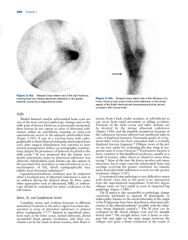
Figure 3.182. Delayed phase lateral view of the right humerus,
showing focal and intense abnormal radiotracer in the greater Figure 3.183. Delayed phase lateral view of the left tarsus of a
tubercle, caused by a suppurative process. horse, showing focal areas of abnormal radiotracer on the dorsal
aspect of the distal intertarsal and tarsometatarsal joints (arrow),
consistent with osteoarthritis.
Stifle
Medial femoral condyle subchondral bone cysts are nature, from a kick, trailer accident, or self‐inflicted as
one of the most common pathologic changes seen in the can occur from rapid movement or falling accidents.
stifle joint of horses. However, as previously mentioned, Fractures of the tuber coxae and tuber ischium can
these lesions do not appear as areas of abnormal radi be detected by the intense abnormal radiotracer
otracer unless an osteoblastic response or juxta‐cyst (Figure 3.186) and the possible asymmetric location of
osteosclerosis occurs in the adjacent subchondral bone the radiotracer between affected and unaffected sides in
(Figure 3.185). A case of a yearling horse with radio cases of displaced fractures. Decreased uptake of a frac
graphically evident bilateral enlarging subchondral bone tured tuber coxae has been associated with a ventrally
13
cysts after surgical debridement was reported to have displaced fracture fragment. Oblique views of the pel
normal postoperative follow‐up scintigraphic examina vis are very useful for evaluating the iliac wing in sus
42
tions, despite the persistence of lameness localized to the pected cases of stress fractures. Trochanteric bursitis is
stifle joints. It was proposed that the lesions were more common in Standardbred racehorses, usually as a
79
mostly osteoclastic; hence no abnormal radiotracer was result of trauma, either direct or related to stress from
56
observed. Subchondral cystic lesions can also appear in racing. Most of the time the lesion involves soft tissue
the proximal tibia secondary to osteochondrosis or as a structures, but in some cases the subchondral bone and
manifestation of OA, which scintigraphically may cartilage overlying the greater trochanter are injured,
exhibit focal abnormal radiotracer. 89 and a focal abnormal radiotracer is seen over the greater
Ligamentous/tendinous avulsions may be suspected trochanter (Figure 3.187).
when a localized area of abnormal radiotracer is seen at Coxofemoral joint pathology is very difficult to assess
an enthesis during the delayed phase. In these cases, with dorsal views due to the great γ‐ray attenuation
other approaches such as ultrasound, MRI, or arthros from the superimposed musculature. Forty‐five degree
copy should be considered for better evaluation of the oblique views are very useful in cases of suspected hip
soft tissues. pathology (Figure 3.188). 13
The SI region is also susceptible to pathologic change
commonly attributed to regional SI desmopathy or
Pelvis, SI, and Coxofemoral Joints arthropathy. Injuries to the sacral tuberosity at the origin
Complete, stress, and avulsion fractures at different of the SI ligaments have been described as abnormal radi
25
anatomical locations in the pelvic region have been doc otracer at the affected enthesis possibly with the tuber
13
umented as areas of abnormal radiotracer in delayed osity being displaced cranially. The SI joint can be
phase scintigraphy. 11,13,38,64 Injuries to superficial struc evaluated on the dorsal oblique view and the straight
tures such as the tuber coxae, ischial tuberosity, dorsal dorsal view. The straight dorsal view is better to com
32
sacrum/tail head, greater trochanter, and third tro pare left and right on the same image; however, the
chanter can be the result of direct trauma, often blunt in oblique view gives a better evaluation of the extent of