
Page 197 - Basic Monitoring in Canine and Feline Emergency Patients
P. 197
usually this number is much less than this (usually
less than 10,000 platelets/hpf) before clinical bleed-
VetBooks.ir ing is noted. In general, the lack of platelets means
that there are not enough cell surfaces for propaga-
tion of coagulation as well as a lack of platelet
granule contents to help in clot formation (see
Section 9.1 for more information).
The most common reasons for thrombocytope-
nia are lack of production of platelets within the
bone marrow, destruction of platelets, or consump-
tion of platelets. Theoretically, any disorder affect-
ing the megakaryocytes can lead to a lack of
production of platelets including neoplastic disor-
ders, immune-mediated destruction of bone mar-
row cells, or other infiltrative disease affecting the
bone marrow. Side effects of various drugs includ-
ing chemotherapeutic agents can also damage the
bone marrow. Destruction of mature platelets is
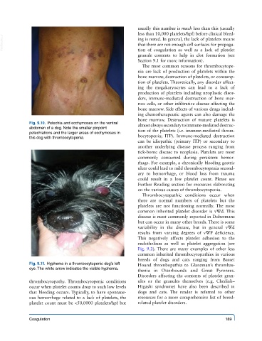
Fig. 9.10. Petechia and ecchymoses on the ventral almost always secondary to immune-mediated destruc-
abdomen of a dog. Note the smaller pinpoint tion of the platelets (i.e. immune-mediated throm-
petechiations and the larger areas of ecchymoses in bocytopenia; ITP). Immune-mediated destruction
this dog with thrombocytopenia.
can be idiopathic (primary ITP) or secondary to
another underlying disease process ranging from
tick-borne disease to neoplasia. Platelets are most
commonly consumed during persistent hemor-
rhage. For example, a chronically bleeding gastric
ulcer could lead to mild thrombocytopenia second-
ary to hemorrhage, or blood loss from trauma
could result in a low platelet count. Please see
Further Reading section for resources elaborating
on the various causes of thrombocytopenia.
Thrombocytopathic conditions occur when
there are normal numbers of platelets but the
platelets are not functioning normally. The most
common inherited platelet disorder is vWd. This
disease is most commonly reported in Dobermans
but can occur in many other breeds. There is some
variability in the disease, but in general vWd
results from varying degrees of vWF deficiency.
This negatively affects platelet adhesion to the
endothelium as well as platelet aggregation (see
Fig. 9.2). There are many examples of other less
common inherited thrombocytopathies in various
breeds of dogs and cats ranging from Basset
Fig. 9.11. Hyphema in a thrombocytopenic dog’s left Hound thrombopathia to Glanzman’s thrombas-
eye. The white arrow indicates the visible hyphema.
thenia in Otterhounds and Great Pyrenees.
Disorders affecting the contents of platelet gran-
thrombocytopathy. Thrombocytopenic conditions ules or the granules themselves (e.g. Chediak–
occur when platelet counts drop to such low levels Higashi syndrome) have also been described in
that bleeding occurs. Typically, to have spontane- dogs and cats. The reader is referred to other
ous hemorrhage related to a lack of platelets, the resources for a more comprehensive list of breed-
platelet count must be <50,0000 platelets/hpf but related platelet disorders.
Coagulation 189