
Page 194 - Basic Monitoring in Canine and Feline Emergency Patients
P. 194
VetBooks.ir
20 m
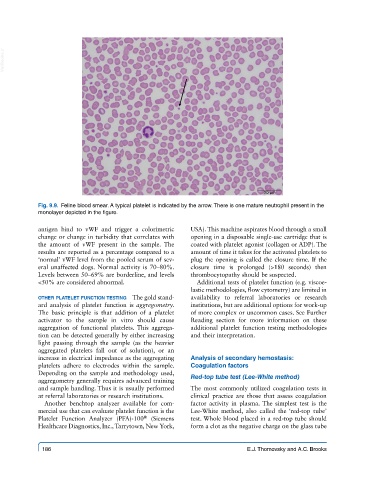
Fig. 9.9. Feline blood smear. A typical platelet is indicated by the arrow. There is one mature neutrophil present in the
monolayer depicted in the figure.
antigen bind to vWF and trigger a colorimetric USA). This machine aspirates blood through a small
change or change in turbidity that correlates with opening in a disposable single-use cartridge that is
the amount of vWF present in the sample. The coated with platelet agonist (collagen or ADP). The
results are reported as a percentage compared to a amount of time it takes for the activated platelets to
‘normal’ vWF level from the pooled serum of sev- plug the opening is called the closure time. If the
eral unaffected dogs. Normal activity is 70–80%. closure time is prolonged (>180 seconds) then
Levels between 50–69% are borderline, and levels thrombocytopathy should be suspected.
<50% are considered abnormal. Additional tests of platelet function (e.g. viscoe-
lastic methodologies, flow cytometry) are limited in
other platelet function testing The gold stand- availability to referral laboratories or research
ard analysis of platelet function is aggregometry. institutions, but are additional options for work-up
The basic principle is that addition of a platelet of more complex or uncommon cases. See Further
activator to the sample in vitro should cause Reading section for more information on these
aggregation of functional platelets. This aggrega- additional platelet function testing methodologies
tion can be detected generally by either increasing and their interpretation.
light passing through the sample (as the heavier
aggregated platelets fall out of solution), or an
increase in electrical impedance as the aggregating Analysis of secondary hemostasis:
platelets adhere to electrodes within the sample. Coagulation factors
Depending on the sample and methodology used,
aggregometry generally requires advanced training Red-top tube test (Lee-White method)
and sample handling. Thus it is usually performed The most commonly utilized coagulation tests in
at referral laboratories or research institutions. clinical practice are those that assess coagulation
Another benchtop analyzer available for com- factor activity in plasma. The simplest test is the
mercial use that can evaluate platelet function is the Lee-White method, also called the ‘red-top tube’
Platelet Function Analyzer (PFA)-100 (Siemens test. Whole blood placed in a red-top tube should
®
Healthcare Diagnostics, Inc., Tarrytown, New York, form a clot as the negative charge on the glass tube
186 E.J. Thomovsky and A.C. Brooks