
Page 192 - Basic Monitoring in Canine and Feline Emergency Patients
P. 192
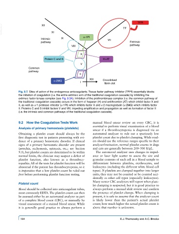
TFPI
  VetBooks.ir                   XI                                         TF
                          XII


                          Intrinsic   IX           α-1PI         VII
                          pathway                                              Extrinsic
                                            VIII                               pathway


                                                 X
                            Protein C and S                    H+AT
                                                 V
                  Common
                  pathway
                                                 II
                                                       XIII
                                 α-2MG                         Crosslinked
                                                 I              fibrin clot

            Fig. 9.7.  Sites of action of the endogenous anticoagulants. Tissue factor pathway inhibitor (TFPI) essentially blocks
            the initiation of coagulation (i.e. the entire extrinsic arm of the traditional coagulation cascade) by inhibiting the
            extrinsic factor tenase complex (see Fig. 9.3A). Inhibition of the prothrombinase complex (i.e. the common pathway of
            the traditional coagulation cascade) occurs in the form of heparan (H) and antithrombin (AT) which inhibit factor X and
            II, as well as α-1 protease inhibitor (α-1PI) which inhibits factor X and α-2 macroglobulin (α-2MG) which inhibits factor
            II. Proteins C and S inhibit factors V and VIII, impeding amplification and propagation as well as formation of factor II
            (i.e. the intrinsic and common pathways of the traditional coagulation cascade).


            9.2  How the Coagulation Tests Work          manual blood smear review on every CBC, it is
                                                         essential to perform visual examination of a blood
            Analysis of primary hemostasis (platelets)
                                                         smear if a thrombocytopenia is diagnosed via an
            Obtaining a platelet count should always be the   automated analyzer to rule out a spuriously low
            first diagnostic test in patients presenting with evi-  platelet count due to platelet clumping. While read-
            dence of a primary hemostatic disorder. If clinical   ers should use the reference ranges specific to their
            signs of a primary hemostatic disorder are present   analyzer/institution, normal platelet counts in dogs
            (petechia, ecchymosis, epistaxis, etc.; see Section   and cats are generally between 200–500 K/μL.
            9.3), but platelet counts are determined to be within   The automated analyzer uses changes in imped-
            normal limits, the clinician may suspect a defect of   ance or laser light scatter to assess the size and
            platelet  function,  also  known  as  a  thrombocy-  granular contents of each cell in a blood sample to
            topathia. All of the tests for platelet function will be   differentiate between platelets, erythrocytes, and
            abnormal if the patient has thrombocytopenia, so it   leukocytes (including the different white blood cell
            is imperative that a low platelet count be ruled out   types). If platelets are clumped together into larger
            first before performing platelet function testing.  units, they may not be counted or be counted acci-
                                                         dentally as other cell types (especially leukocytes).
                                                         Many newer CBC analyzers will report when plate-
            Platelet count
                                                         let clumping is suspected, but it is good practice to
            Blood should be collected into anticoagulant tubes,   always perform a manual slide review and confirm
            most commonly EDTA. The platelet count can then   the presence of platelet clumps. When clumping is
            be assessed either by an automated analyzer as part   present, it is safe to assume that the analyzer count
            of a complete blood count (CBC), or manually by   is likely lower than the patient’s actual platelet
            visual assessment of a stained blood smear. While   count; how much higher the actual platelet count is
            it  is generally good practice to always perform a   above that number is unknown.


             184                                                         E.J. Thomovsky and A.C. Brooks