
Page 187 - Basic Monitoring in Canine and Feline Emergency Patients
P. 187
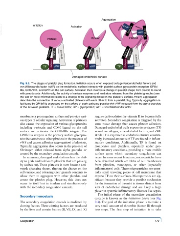
Aggregation
  VetBooks.ir       Initiation             Activation                    Platelet  vWF GpIIb/IIIa  vWF Platelet  GPIIb/IIIa


                             Platelet                  Platelet               Platelet
                                                                                       Platelet

                              GPIIb/IIIa  GPVI  GPIb/V/IX  GpIIb/IIIa  GpVI  GPIb/V/IX  GpIIb/IIIa  GpVI


                                Collagen             Collagen               Collagen  GPIb/V/IX
                           TF    vWF            TF    vWF              TF    vWF


                                            Damaged endothelial surface
             Fig. 9.2.  The stages of platelet plug formation. Initiation occurs when exposed collagen/subendothelial factors and
             von Willebrand’s factor (vWF) on the endothelial surface interacts with platelet surface glycoprotein receptors GPIIb/
             IIIa, GPIb/V/IX, and GPVI on the cell surface. Activation then involves a change in platelet shape from discoid to round
             with pseudopods. Additionally, the activity of various enzymes and mediators released from the platelet granules (see
             the text for more information) leads to a change in the signaling milieu on the platelet’s surface. Finally, aggregation
             describes the connection of various activated platelets with each other to form a platelet plug. Typically, aggregation is
             facilitated by GPIIb/IIIa expressed on the surface of each activated platelet with vWF released from the alpha granules
             of the activated platelets. TF = tissue factor; GP = glycoprotein; vWF = von Willebrand’s factor.

             membrane a procoagulant surface and provide vari-  require carboxylation by vitamin K to become fully
             ous types of cellular signaling. Activation of platelets   activated. Secondary coagulation is triggered by the
             also causes the expression of various glycoproteins   same tissue damage that causes platelet adhesion.
             including p-selectin and CD40 ligand on the cell   Damaged endothelial walls expose tissue factor (TF)
             surface and activates the GPIIb/IIIa integrin.  The   as well as collagen, subendothelial factors, and vWF.
             GPIIb/IIIa integrin is the primary surface glycopro-  While TF is expressed in endothelial tissues constitu-
             tein that attaches to other platelets in the presence of   tively, increased amounts of TF are found in inflam-
             vWF and causes adhesion (aggregation) of platelets.   matory conditions.  Additionally,  TF  is found on
             Typically, aggregation also occurs in the presence of   monocytes and platelets, especially under pro-
             fibrinogen either released from alpha granules or   inflammatory conditions, providing a more mobile
             created by the secondary coagulation cascade.  surface upon which secondary coagulation can
               In summary, damaged endothelium has the abil-  occur. In more recent literature, microparticles have
             ity to grab and hold onto platelets that are passing   been described which are blebs of cell membranes
             by (adhesion). These platelets in turn become acti-  from platelets, monocytes, or other (especially
             vated: changing shape, altering the charge on the   inflammatory) cells. These microparticles are essen-
             cell surface, and releasing their granule contents to   tially small traveling pieces of cell membrane that
             allow them to aggregate with other platelets and   express TF on their surfaces. Microparticles are sig-
             create  the  platelet  plug.  However, this  does  not   nificant because they provide a possible mechanism
             occur by itself but in tandem and simultaneously   for the formation of thrombi in tissues distant from
             with the secondary coagulation cascade.     sites of endothelial damage and are likely a large
                                                         player in systemic inflammatory illnesses like sepsis.
                                                           The initial phase of the secondary coagulation
             Secondary hemostasis
                                                         cascade is known as the initiation phase (see Fig.
             The secondary coagulation cascade is mediated by   9.3). The goal of the initiation phase is to make a
             clotting factors. These clotting factors are produced   very small amount of thrombin (factor II) through
             by the liver and certain factors (II, VII, IX, and X)   two steps.  The first step of initiation is to take


             Coagulation                                                                     179