
Page 2166 - Cote clinical veterinary advisor dogs and cats 4th
P. 2166
Chest Tube Placement 1083
injecting, aspirate back to determine that a guard to prevent the tube from entering • Bandage to secure tube(s)
the thorax excessively.
needle is not in the intercostal artery or vein. • Tube is slid off the trocar/stylet cranially and • Pain management: injectable opioids versus
VetBooks.ir ventilation while inserting tube into thorax ventrally along chest wall, then connected can be used for optimal pain control)
intrathoracic bupivacaine (both modalities
• If the patient is anesthetized, stop manual
and secured as described previously.
to deflate lungs and decrease risk of lung
○ Bupivacaine can be given (1.5 mg/kg)
trauma.
tube q 6-8h.
recommended in dogs only in emergency
• With the skin still drawn cranially, hemostats • This very rapid placement technique is diluted 1 : 1 with 0.9% NaCl through
are used for bluntly dissecting vertically into situations because of increased risk of iat- • Continuous monitoring as long as the chest
the pleural space, spreading the jaws wide rogenic trauma and is never recommended tube is in place because of the risks of discon-
enough for the tube to snugly fit through the in cats because of their very compliant chest nection and development of pneumothorax
opening. The tube is inserted through the walls.
jaws of the hemostat and advanced cranially Alternate method: insertion of guide wire–based Alternatives and Their
and ventrally along the chest wall, making chest tube Relative Merits
sure that all side holes in the tube are well • Local anesthesia (± sedation if needed) Repeat thoracocentesis: may become difficult to
within the thorax. The distance the tube is • Seldinger technique: as for multilumen manage animal if pleural evacuation is needed
advanced can be measured with the stylet. jugular catheters (p. 1123) frequently Procedures and Techniques
The tip of the tube should be at the second • An assistant pulls the skin cranially several
or third ICS. centimeters and holds it in place while the Pearls
• Connect tube to a 3-way stopcock and tube is inserted. This creates a SQ tunnel The risk of impaling lung or heart can be
injection caps or a pleural drainage system. once released. An 18- or 14-gauge catheter minimized with the trocar technique if the
• Secure to skin with a purse-string suture introducer is placed into the pleural space operator holds the distal portion of the tube
around the tube at the entry site and a through the seventh or eighth ICS. and trocar in a clenched fist, with 1-2 inches of
Chinese finger trap suture pattern (with or • A 60-cm guide wire is inserted through the tube and trocar protruding, to prevent excessive
without tissue glue to affix the suture to the catheter, leaving ≈20 cm or more outside the penetration into the thorax.
tube) to reduce sliding of the tube. thorax.
• Place sterile dressing and light bandage. • The catheter introducer is removed, leaving SUGGESTED READING
• Tube connection sites can be secured with the guide wire in place. Lombardi R, et al: Pleural space drainage. In Burkitt
orthopedic wire in figure-eight patterns or • The 14-gauge catheter is inserted into the Creedon JM, et al, editors: Advanced monitoring
plastic zip ties. pleural space over the guide wire, the guide and procedures for small animal emergency and
Alternate method, trocar technique: wire is removed, and the catheter is sutured critical care, West Sussex, UK, 2012, Wiley-
• Sedation/anesthesia, initial prep as previously in place. Blackwell, pp 378-392.
described • Small-diameter and flexible material make it
• Tube and trocar/stylet within it are tunneled well tolerated, especially in cats, small dogs, RELATED CLIENT EDUCATION
SQ by 2-3 rib spaces and then positioned and animals with pneumothorax. SHEETS
perpendicular to the chest wall and grasped • Can become obstructed if effusion is thick
tightly 1-2 inches from distal tip. or kink if placed in animals with a thick Consent to Perform General Anesthesia
• Top of the tube will be hit bluntly with the chest wall (large or obese dogs) Consent to Perform Thoracocentesis
palm of the other hand, popping the tube
through the thoracic wall and into pleural Postprocedure AUTHOR: Lori S. Waddell, DVM, DACVECC
EDITORS: Leah A. Cohn, DVM, PhD, DACVIM; Mark S.
space; the other hand, grasping tube and • Thoracic radiographs (lateral and ventrodorsal Thompson, DVM, DABVP
trocar 1-2 inches from the distal tip, acts as or dorsoventral) to check tube(s) placement
12 34 56 78 9101112
A C
B
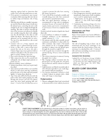
CHEST TUBE PLACEMENT A, For placement of a chest tube, animal is in lateral recumbency, and an incision is
made in the skin at least two intercostal spaces (ICSs) caudal to planned site of entry into thorax. B, Chest tube and
trocar within it are tunneled together SQ to the appropriate ICS, and chest is entered with the tube. C, Tube is then
advanced off the trocar (small arrows) such that all side holes of the chest tube are within the pleural space and trocar
has not advanced farther into chest. Trocar is then withdrawn (large arrow), and chest tube is capped and secured.
www.ExpertConsult.com