
Page 2164 - Cote clinical veterinary advisor dogs and cats 4th
P. 2164
Cerebrospinal Fluid Collection 1081
• While maintaining a grip on the hub of the case, the needle is removed and discarded, • Sterile gloves are worn, but the field is not
usually draped.
needle with the right hand, the clinician gloves are changed, the surgical prep is • The animal is positioned in lateral recum-
VetBooks.ir • The stylet is withdrawn with the right hand, again. NOTE: If blood is encountered, bency with the hindlimbs and lumbar spine
uses the left hand to grasp the needle near
repeated, and the procedure is started
its insertion into the skin.
it is typically from a location outside the
flexed (tucked posture).
and the needle hub is examined for CSF
flow. subarachnoid space and does not limit the Landmarking and puncture:
• The needle enters the L5-L6, or L6-L7
ability to obtain an uncontaminated sample
• If no CSF is present, the needle is first when the procedure is repeated. interspace in dogs or the L7-S1 interspace
withdrawn a few millimeters while watching • After CSF begins to drip from the hub, it in cats.
for the appearance of CSF to ensure the is collected into two plain, sterile, red-top • Except in very small or thin animals, the L6
needle has not been inserted too deeply tubes. Ideally, an assistant collects the sample, dorsal spine is typically the most caudal and
(into nervous tissue), and then the needle and the examiner does not release the needle can be palpated just cranial to the wings of
is advanced. The stylet need not be replaced during collection. the ilium.
at this point. (Although advancing the • In general, 1 mL of CSF is required for • If the L7 dorsal spine is palpable, it is
needle without the stylet creates a slight assessment of the white blood cell (WBC) usually much smaller than that of L6 and
risk of creating a tissue plug in the needle, count, protein concentration, and cytologic lies between the wings of the ilium. Procedures and Techniques
it decreases the risk of advancing the needle examination (1 mL/5 kg of body weight can • Radiographs can demonstrate individual
too deeply because CSF appears as soon as be obtained safely). differences in anatomy.
the subarachnoid space is entered.) • If the animal weighs > 5 kg, additional • The needle is inserted into the skin just
• As the needle is advanced, one of three things CSF can and should be collected for caudolaterally to the caudal dorsal spine of
happens: possible culture or ancillary diagnostics, the space to be entered (i.e., caudolaterally
○ CSF appears in the hub of the needle such as infectious titers, flow cytometry, or to L6 for puncture between L5 and L6).
and begins to drip out. The needle should immunoglobulin indices. • The needle is directed craniomedially to
then be advanced 1 mm farther to place • After the appropriate amount of CSF has puncture the interarcuate space between
the entire bevel in the cerebellomedullary been collected, the needle is removed. the vertebrae.
cistern, and the needle is grasped firmly • NOTE: If during the procedure the heart rate • If bone is encountered, the needle is redi-
against the skin. The slight advancement acutely decreases or fluid streams rather than rected (usually cranially) until the space is
and firm hold on the needle reduce the risk drips from the hub when the subarachnoid identified.
of blood contamination from meningeal space is entered, the needle should be • If the needle is inserted to the hub and no
vessels. removed and the procedure aborted. bone is encountered, the needle is too short
○ The needle hits bone. This is usually the or directed too far laterally.
occipital bone, and the needle should be LUMBAR APPROACH • Fluid can be collected from the dorsal
redirected caudally to enter the cerebel- • General anesthesia and intubation subarachnoid space, but more often the
lomedullary cistern; if it strikes the atlas, • Preparation and positioning (for centesis from needle is passed through the nervous tissue
the needle must be redirected cranially. the lumbar subarachnoid space) is technically to the floor of the vertebral canal.
The needle may be walked along the bone more difficult, yields less fluid, and is more • The stylet is removed, and if CSF is not
to find the cistern, bearing in mind that likely to involve blood contamination but recovered, the needle is withdrawn slightly
it will be only millimeters deeper, but this is more sensitive for focal thoracolumbar to enter the ventral subarachnoid space while
may result in blood contamination from lesions. the examiner watches for the appearance of
trauma to the periosteum. • The skin is shaved and aseptically prepared fluid in the hub of the needle.
○ Frank blood appears in the hub. This most over the dorsal midline from L3 cranially • CSF typically flows much more slowly from
likely indicates that the needle is off midline to the midsacrum caudally and the wings this site compared with the cerebellomedul-
and has punctured a venous sinus. In this of the ilium laterally. lary cistern.
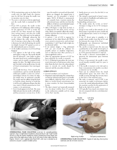
Needle Left wing
insertion site of atlas
CEREBROSPINAL FLUID COLLECTION Landmarks for cerebellomedullary
approach. Dog is in right lateral recumbency, head pointing to right. Right-handed
clinician is using left hand to identify the occipital protuberance (index finger) and
wings of the atlas (right wing of atlas: thumb; left wing of atlas: middle and ring Right wing of atlas Occipital protuberance
fingers). Patient’s neck is appropriately flexed (90°), and nose is elevated by an CEREBROSPINAL FLUID COLLECTION Diagram of same dog, showing bony
assistant so muzzle is parallel to table surface. landmarks and site of needle insertion.
www.ExpertConsult.com