Page 2373 - Cote clinical veterinary advisor dogs and cats 4th
P. 2373
Transtracheal Wash 1172.e5


            Transtracheal Wash
  VetBooks.ir                                  Anticipated Time


           Difficulty Level: ♦♦
                                                                                    further vertical (45° toward ceiling); if one
                                               About 15-20 minutes                •  Head  and  neck  elevated  to  horizontal  or
           Synonyms                                                                 side of the lungs is of significantly greater
           TTW, transtracheal aspiration (TA), tracheal   Preparation: Important    interest, have it be the dependent (down)
           lavage                              Checkpoints                          side, using lateral recumbency
                                               •  Recent thoracic radiographs     •  Palpate the small cricothyroid membrane at
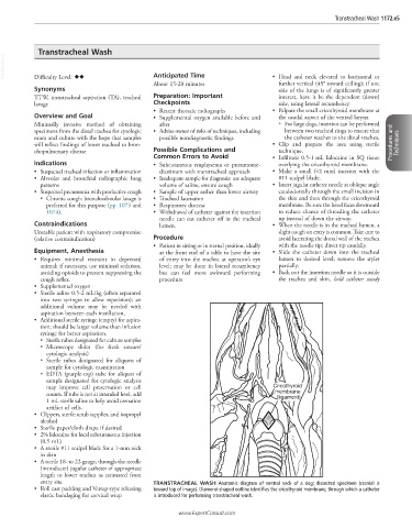
           Overview and Goal                   •  Supplemental oxygen available before and   the caudal aspect of the ventral larynx
           Minimally  invasive  method  of  obtaining   after                       ○   For large dogs, insertion can be performed
           specimens from the distal trachea for cytologic   •  Advise owner of risks of techniques, including   between two tracheal rings to ensure that
           exam and culture with the hope that samples   possible nondiagnostic findings.  the catheter reaches to the distal trachea.
           will reflect findings of lower tracheal to bron-                       •  Clip  and  prepare  the  area  using  sterile   Procedures and   Techniques
           chopulmonary disease                Possible Complications and           technique.
                                               Common Errors to Avoid             •  Infiltrate 0.5-1 mL lidocaine in SQ tissue
           Indications                         •  Subcutaneous  emphysema  or  pneumome-  overlying the cricothyroid membrane.
           •  Suspected tracheal infection or inflammation  diastinum with transtracheal approach  •  Make  a  small  (<1 mm)  incision  with  the
           •  Alveolar  and  bronchial  radiographic  lung   •  Inadequate sample for diagnosis: use adequate   #11 scalpel blade.
             patterns                           volume of saline, ensure cough    •  Insert jugular catheter needle at oblique angle
           •  Suspected pneumonia with productive cough  •  Sample of upper rather than lower airway  caudodorsally through the small incision in
             ○   Chronic cough: bronchoalveolar lavage is   •  Tracheal laceration  the skin and then through the cricothyroid
               preferred for this purpose (pp. 1073 and   •  Respiratory distress   membrane. Be sure the bevel faces downward
               1074).                          •  Withdrawal of catheter against the insertion   to reduce chance of threading the catheter
                                                needle can cut catheter off in the tracheal   up instead of down the airway.
           Contraindications                    lumen.                            •  When the needle is in the tracheal lumen, a
           Unstable patient with respiratory compromise                             slight cough on entry is common. Take care to
           (relative contraindication)         Procedure                            avoid lacerating the dorsal wall of the trachea
                                               •  Patient in sitting or in sternal position, ideally   with the needle tip; direct tip caudally.
           Equipment, Anesthesia                at the front end of a table to have the site   •  Slide  the  catheter  down  into  the  tracheal
           •  Requires  minimal  restraint  in  depressed   of entry into the trachea at operator’s eye   lumen to desired level; remove the stylet
             animal; if necessary, use minimal sedation,   level;  may  be  done  in  lateral  recumbency   partially.
             avoiding opioids to prevent suppressing the   but  can  feel  more  awkward  performing    •  Back out the insertion needle so it is outside
             cough reflex.                      procedure                           the  trachea  and  skin,  hold  catheter  steady
           •  Supplemental oxygen
           •  Sterile saline 0.5-2 mL/kg (often separated
             into two syringes to allow repetition); an
             additional  volume  may be needed  with
             aspiration between each instillation,
           •  Additional sterile syringe (empty) for aspira-
             tion; should be larger volume than infusion
             syringe for better aspiration.
             ○   Sterile tubes designated for culture samples
             ○   Microscope  slides  (for  fresh  smears/
               cytologic analysis)
             ○   Sterile  tubes  designated  for  aliquots  of
               sample for cytologic examination
             ○   EDTA  (purple-top)  tube  for  aliquot  of
               sample designated for cytologic analysis
               may improve cell preservation or cell                              Cricothyroid
                                                                                         d
               counts. If tube is not at intended level, add                       membrane
                                                                                   (ligament)
               1 mL sterile saline to help avoid crenation
               artifact of cells.
           •  Clippers, sterile scrub supplies, and isopropyl
             alcohol
           •  Sterile paper/cloth drape if desired
           •  2% lidocaine for local subcutaneous injection
             (0.5 mL)
           •  A sterile #11 scalpel blade for a 1-mm nick
             in skin
           •  A sterile 18- to 22-gauge, through-the-needle
             (introducer) jugular catheter of appropriate
             length to lower trachea as estimated from
             entry site                        TRANSTRACHEAL  WASH  Anatomic diagram of ventral neck of a dog; dissected specimen (cranial is
           •  Roll cast padding and Vetrap-type releasing   toward top of image). Diamond-shaped outline identifies the cricothyroid membrane, through which a catheter
             elastic bandaging for cervical wrap  is introduced for performing transtracheal wash.

                                                      www.ExpertConsult.com

