
Page 2376 - Cote clinical veterinary advisor dogs and cats 4th
P. 2376
1174 Ureteral Occlusion, Subcutaneous Ureteral Bypass
Ureteral Occlusion, Subcutaneous Ureteral Bypass Client Education
Sheet
VetBooks.ir
ureteral calculi in which only a short segment
Difficulty level: ♦♦♦
of proximal ureter remains. • If a urinary tract infection is present, begin
appropriate antibiotics (pp. 232 and 849)
Synonym prior to surgery, ideally 48-72 hours prior.
SUB Contraindications
• Although not an absolute contraindication, Possible Complications and
Overview and Goals mineralization of an SUB device in dogs Common Errors to Avoid
• Ureteral obstructions are a major clinical is more common than mineralization of • Technical error is avoided by proper
problem in cats and dogs, and are most ureteral stents resulting in re-occlusion. training.
commonly associated with ureteroliths, Therefore, stent placement usually is preferred • Mineralization of the device (24% of cats) is
ureteral strictures, or trigonal neoplasia of in dogs. minimized by use of a tetra-sodium EDTA
the ureterovesicular junction. Traditional • A small-diameter renal pelvis (<8 mm) does flush q 3 months. Despite mineralization
surgical treatment options are more invasive require the nephrostomy tube to be modified of the device, only 12% of cats needed an
and are reported to have greater morbidity as a ureterostomy tube, but this does not SUB exchange due to development of ureteral
than the newer interventional options such preclude placement of the SUB device. The patency and failure or ureteral re-obstruction.
as ureteral stent placement or subcutaneous author has successfully placed SUB devices Mineralization is more likely in dogs than
ureteral bypass (SUB) device. in cats with a renal pelvis under 3 mm in cats after SUB placement.
• In the author’s experience, use of the SUB diameter. • Chronic urinary tract infections (8% of
device is preferred for cats over placement of cats) are minimized by use of a tetra-sodium
ureteral stents, and it can be used in specific Equipment, Anesthesia EDTA flush q 3 months and amenable to use
cases in dogs. In the past 9 years, the author • General anesthesia is required. of antimicrobial drugs chosen on the basis
has placed > 500 SUB devices with good to • SUB device: a polyurethane catheter (6.5 Fr) of urine culture and susceptibility testing. It
excellent results (especially in cats) in the that is composed of a locking-loop nephros- is usually unnecessary to treat asymptomatic
short and long term. tomy and cystostomy catheter and a metallic bacteriuria associated with Enterococcus spp
shunting port that connects the catheters (most common potential pathogen isolated
Indications subcutaneously, allowing for drainage and from these cats).
• Cats with any cause of a ureteral obstruc- system flushing/sampling • Dysuria is rare (<5%) after SUB device
tion (e.g., ureteroliths [p. 1008], strictures, • A traditional fluoroscopic C-arm is sufficient placement.
tumors, extraluminal ureteral compression). for visualization during ureteral interventions.
A SUB device can be predictably placed in • Various guide wires and catheters are needed Procedure
cats regardless of the cat’s size or severity of for each procedure. The procedure should be done by trained
obstruction. interventionalists using fluoroscopic guidance
• In dogs, SUB can be considered instead Anticipated Time and surgical assistance.
of ureteral stent placement if there is a In experienced hands, an SUB device can be
ureteral stent reaction, ureteral surgery is too placed, on average, in 45 minutes unilater- Postprocedure
complicated, or a radical surgery is being ally, and 60 minutes bilaterally. Typically, an • Esophagostomy tube (p. 1106) is usually
considered for removal of transitional cell esophagostomy tube is placed after surgery placed to ensure appropriate nutrition,
carcinoma. is complete, which can add an additional 10 medications, and hydration, ultimately
• Interventional procedures are especially minutes to the procedure time. shortening hospitalization time (median,
helpful for cats that could benefit from 4 days).
reduced anesthesia time or cases that could be Preparation: Important • Flush each SUB device regularly, typically at
challenging using traditional ureteral surgery, Checkpoints 1 week, 1 month, and then every 3 months
such as proximal ureteral obstructions • Appropriate hydration prior to anesthesia thereafter using the tetra-sodium EDTA
(stones/stricture), numerous ureterolithiasis should be a goal. Many patients are over- solution to help avoid mineralization and
requiring multiple ureterotomies, extensive hydrated and minimal additional fluids are chronic urinary tract infections.
ureteral injury, or trauma associated with needed during surgery. • Urinary tract ultrasounds should be done
every few months for all patients with a
history of a ureteral obstruction due to the
high risk of the contralateral kidney being
obstructed.
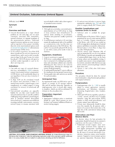
ShuntingPort with 2 catheter outlets
Alternatives and Their
Relative Merits
The decision about the best procedure for
Flow is from the kidney each patient should be based on the operator’s
through the shuntingport
and into the bladder - experience and the cause of obstruction.
Locking loop
kidney catheter bypassing the ureter • Traditional ureteral surgery: more inva-
with marker band sive, longer anesthesia times, reported to
have higher perioperative morbidity and
A B mortality rates, and not readily applicable
Fenestrated & cuffed bladder catheter for all causes of ureteral obstruction (e.g.,
URETERAL OCCLUSION, SUBCUTANEOUS URETERAL BYPASS A, A lateral fluoroscopy image of a stricture, numerous stones, stone location,
SUB device after implantation. Notice the locking-loop nephrostomy tube in the renal pelvis and the cystostomy tumors). Surgical or interventional options
tube in the urinary bladder. Both are hooked to a subcutaneous shunting port. B, The SUB device. are operator dependent. Decisions about
www.ExpertConsult.com