
Page 2379 - Cote clinical veterinary advisor dogs and cats 4th
P. 2379
1175.e2 Ureteral Stent Placement
Ureteral Stent Placement Client Education
Sheet
VetBooks.ir Equipment, Anesthesia
Difficulty level: ♦♦♦
• General anesthesia is required. recumbency. For male dogs, it can be done using
flexible retrograde cystoscopy, retrograde peri-
Synonyms • Ureteral stents: double-pigtail, multifenes- neal urethral access, or percutaneous antegrade
Ureteral stent, double-pigtail ureteral stent trated design with one loop curled in the access from the bladder apex. They all allow
renal pelvis, the shaft traveling in the ureteral retrograde ureteral access using fluoroscopic
Overview and Goal lumen, and the other loop curled in the guidance. Various guide wires and catheters
Ureteral obstructions are a major clinical urinary bladder lumen are needed for each procedure.
problem in dogs and cats and are most com- • Various rigid and flexible endoscopes
monly associated with ureteroliths, ureteral • Fluoroscopy equipment Postprocedure
strictures, and trigonal neoplasia of the Patients are often discharged the same
ureterovesicular junction (UVJ). Traditional Anticipated Time day as the procedure, and the recovery is
surgical treatment options are more invasive 15-45 minutes when done endoscopically by minimal. Six weeks of an appropriate antibi-
and have greater morbidity than interventional trained operators otic should be used if the urine is infected
options such as ureteral stent placement or (≈60%).
subcutaneous ureteral bypass (SUB [p. 1174]) Preparation: Important
device placement. Interventional options are Checkpoints Alternatives and Their
preferred when possible. The goals of ureteral • Appropriate hydration before anesthesia Relative Merits
stenting, typically reserved for canine patients, • If a urinary tract infection is present, begin The decision about the best procedure for
are fourfold: appropriate antibiotics (pp. 232 and 849). each patient should be based on the operator’s
• Bypass a ureteral obstruction in order to experience and the cause of obstruction.
divert urine from the renal pelvis into the Possible Complications and • Traditional ureteral surgery: more invasive,
urinary bladder Common Errors to Avoid time consuming, liable to greater morbidity
• Encourage passive ureteral dilation to prevent Complications associated with cystoscopy and than interventional procedures, but surgical
re-obstruction, encourage stone passage, or ureteral manipulation are rare when procedures options may be available in areas where there
aid in future ureteroscopy are performed by trained interventionists. The is limited or no access to trained veterinary
• Facilitate surgery of the ureter (especially most important long-term risks are ureteral stent specialty care
during a ureteral resection and anastomosis) reaction resulting in persistent ureteral obstruc- • SUB placement: preferred for cats, applicable
and prevent postoperative leakage and tion, stent migration, or chronic urinary tract for dogs
edema infections (usually amenable to antimicrobial
• Aid in extracorporeal shockwave lithotripsy treatment). Pearls
for complex nephroliths to avoid obstructive • No preoperative imaging or biochemical
ureterolith development Procedure data can predict the overall outcome or renal
Ureteral stents can remain in place for years and This procedure should be done by trained function, but prognosis for long-term survival
are considered a long-term treatment option for interventionalists using endoscopic/fluoroscopic in dogs with a benign cause of ureteral
various causes of ureteral obstructions. guidance. In female dogs, it is typically done obstruction treated with stent placement is
using standard retrograde cystoscopy in dorsal excellent.
Indications
• Any cause of a ureteral obstruction, including
stones, strictures, tumors, or extraluminal
compressions in dogs
• Interventional procedures are especially
helpful for dogs that could benefit from
reduced anesthesia time or cases that could be
challenging using traditional ureteral surgery,
such as proximal ureteral obstructions
(stones/stricture), numerous ureterolithiasis
requiring multiple ureterotomies, extensive
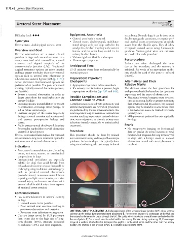
ureteral injury, and trauma associated with A B
ureteral calculi in which only a short segment
of proximal ureter remains.
Contraindications
• Few contraindications to ureteral stenting
in dogs
○ Ureteral access is not possible.
○ Prior ureteral stent reaction resulting in C D E
persistent obstruction and ureteritis
○ Recurrent stent infections or migration URETERAL STENT PLACEMENT A, Endoscopic image of the ureterovesicular junction (UVJ) with a ureteral
• Cats are better served by SUB placement catheter up the orifice during ureteral stent placement. B, Fluoroscopic image of a cystoscope at the UVJ and
the ureteral catheter up the ureter through the UVJ. The guide wire is inside the ureteral lumen and coiled within
than stents due to the high risk of long- the renal pelvis. C, The ureteral stent is coiled within the urinary bladder during cystoscopy. D, Fluoroscopic
term dysuria (38%), stricture associated image of a ureteral stent after it is deployed. One pigtail is in the renal pelvis, and the other is in the urinary
re-occlusion (19%), and stent migration. bladder; the shaft is in the ureteral lumen. E, A double-pigtail ureteral stent.
www.ExpertConsult.com