
Page 2419 - Cote clinical veterinary advisor dogs and cats 4th
P. 2419
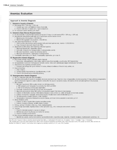
1198.e2  Anemias: Evaluation


            Anemias: Evaluation
  VetBooks.ir  Approach to Anemia Diagnosis


            I.  Determine Severity of Anemia
               A.  Mild anemia (PCV > 30% dog, > 20% cat)
                1.  Consider age, breed, and statistical chance of normality.
                2.  Check for laboratory or sample error; repeat venipuncture.
               B.  For moderate to severe anemia, go to step II.
            II.  Determine Bone Marrow Responsiveness
               A.  No reticulocytosis or polychromasia expected during first 2-3 days or in mild anemia (PCV > 30% dog, > 20% cat).
               B.  Reticulocytosis and polychromasia peak at 4-5 days if bone marrow function normal.
                1.  Marked canine reticulocytosis > 500,000/mcL
                2.  Marked feline aggregate reticulocytosis > 200,000/mcL
               C.  Later-stage responsiveness at 7-14 days
                1.  Feline punctate reticulocytosis (rarely evaluated with automated systems now), marked > 1,500,000/mcL
                2.  Dogs: increase in macrocytic hypochromic RBCs
               D.  Classification by RBC indices and hematology instrument graphics
                1.  Macrocytic hypochromic: regenerative anemia
                2.  Normocytic normochromic: nonregenerative or preregenerative anemia
                3.  Microcytic hypochromic: usually iron-deficiency anemia
                4.  Macrocytic normochromic: regenerative or nonregenerative
               E.  If adequately regenerative, go to step III; if inadequately regenerative, go to step IV.
            III. Regenerative Anemia Diagnosis
               A.  Blood smear analysis critical in hemolytic anemia diagnosis
                1.  Spherocytes, autoagglutination, Heinz bodies, polychromasia, blood parasites, eccentrocytes, RBC fragmentation
               B.  Hemoglobinuria is best proof of intravascular hemolytic anemia. Icterus and splenomegaly suggest extravascular hemolysis.
               C.  Internal blood loss resembles hemolytic anemia.
                1.  Document hemorrhage with gross evidence of melena, ultrasound evidence of blood in body cavities, etc.
               D.  External blood loss
                1.  Often in history
                2.  Tendency toward hypoproteinemia, hypoalbuminemia, or both
                3.  Check for thrombocytopenia or bleeding tendency.
            IV.  Nonregenerative Anemia Diagnosis
               A.  Way to a diagnosis varies with case presentation.
               B.  Use history and severity of anemia to reevaluate reticulocyte numbers to see if anemia is truly nonregenerative; duration exceeding 3-4 days excludes preregenerative
                anemia; reticulocyte response is weak or absent 2 weeks after the cause of an anemia ceases; mild anemia will not stimulate significant reticulocytosis.
               C.  CBC evaluation
                1.  Microcytic hypochromic RBCs usually indicate iron-deficiency anemia
                   a.  RBC cytograms and histograms more sensitive than MCV and MCHC
                   b.  Half of iron-deficiency anemia cases regenerative
                2.  Normocytic normochromic anemia most common but nonspecific
                3.  Macrocytic normochromic feline RBCs without reticulocytosis suggest FeLV-induced myelodysplasia
                4.  Evidence of inflammation; anemia of inflammatory diseases is very common (i.e., mild, normocytic normochromic anemia).
                5.  Evidence of leukemia or dysplastic hematopoiesis usually indicates bone marrow involvement; go to H.
                6.  Thrombocytopenia; consider Ehrlichia or other infections.
                7.  Pancytopenia or bicytopenia indicates bone marrow disease, and bone marrow evaluation is warranted; go to H.
               D.  Clinical chemistry profile
                1.  Evidence of renal or hepatic failure causing secondary anemia
                2.  Evidence of systemic diseases; variable causes of anemia
               E.  Virology, serology if infection is likely (e.g., fever, lymphadenopathy)
               F.  Endocrinologic examination; hypothyroidism or other dysfunction (e.g., mild, normocytic normochromic anemia)
               G.  Toxicosis
                1.  Check for testicular neoplasm or access to estrogen.
                2.  Withhold any current drug therapy and monitor for recovery.
                3.  Check for toxicants in environment.
               H.  Bone marrow examination reveals many diagnoses.
                1.  Myelofibrosis, aplastic anemia, bone marrow necrosis/inflammation, dyserythropoiesis, leukemia, metastatic neoplasia, myelodysplastic syndromes, etc.
           NOTE: The unit of measure of micron, sometimes denoted by the Greek letter μ, is abbreviated in this text by mc. For example, 1 mcL = 1 microliter; 1 mcg = 1 microgram.
           CBC, Complete blood count; FeLV, feline leukemia virus; MCHC, mean corpuscular hemoglobin concentration; MCV, mean corpuscular volume; PCV, packed cell volume; RBC, red blood cell.
           Modified from Willard M, Tvedten H: Small animal clinical diagnosis by laboratory methods, ed 5, St. Louis, 2012, Saunders.


                                                     www.ExpertConsult.com