
Page 2508 - Cote clinical veterinary advisor dogs and cats 4th
P. 2508
Lymphadenopathy Lymphoma Staging Classification 1251
Lymphadenopathy
VetBooks.ir Differential Diagnosis Key Features
Generalized Lymphadenopathy
Reactive:
• Infectious (incomplete list) • The majority of these conditions have concurrent supportive findings:
• Rickettsial infection • Thrombocytopenia, leukopenia, nonregenerative anemia, hyperglobulinemia
• Leishmaniasis • Endemic areas, amastigotes may be detected in lymph node aspirates
• Feline leukemia virus, feline immunodeficiency virus • Leukopenia, nonregenerative anemia; point-of-care testing positive
• Systemic mycoses (histoplasmosis, blastomycosis, cryptococcosis, • Endemic areas, immunosuppressed patients, localized fungal infection with sudden
sporotrichosis, coccidioidomycosis, aspergillosis) deterioration in condition
• External parasites • Skin scrape for Sarcoptes scabiei, Demodex sp, etc.
• Generalized dermatopathy • Apparent on examination
• Noninfectious/inflammatory • May occur with recent trauma or autoimmune-related disorders
Neoplasia:
• Lymphoma, systemic mastocytosis, leukemia, multiple myeloma • Fine-needle aspirate of the popliteal or superficial cervical (prescapular) lymph nodes is
recommended; certain neoplasms (lymphoma, myeloma notably) may be associated with
hypercalcemia, hyperglobulinemia, thrombocytopenia
Nonspecific Hyperplasia • Cause may be undetermined; associated with retrovirus infections in cats and
leishmaniasis in dogs
Solitary or Regional Lymphadenopathy
Superficial:
• Inflammatory conditions of the drained region resulting in lymphadenitis • If only one lymph node is affected, carefully examine the drainage area of the lymph node
(e.g., abscessation, wounds, tick bites, dermatitis, periodontal disease) for the underlying cause (p. 598) Differentials, Lists, and Mnemonics
• Metastatic neoplasia
Deep (Visceral):
• Systemic mycoses • Associated with (often marked) systemic illness
• Metastatic neoplasia • Identify primary neoplasm via physical exam, ultrasound, radiography. Fine-needle aspirate
or biopsy required for final diagnosis
• Inflammatory conditions of the drained region (e.g., enteritis, hepatitis) • Clinical, biochemistry, and/or imaging signs often supportive of primary problem
Reproduced from the third edition in modified form.
THIRD EDITION AUTHOR: Paolo Pazzi, BVSc, MMedVet
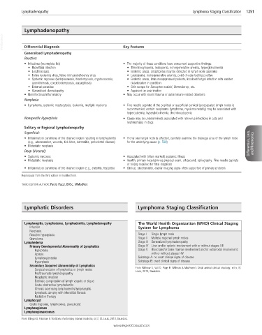
Lymphatic Disorders Lymphoma Staging Classification
Lymphangitis, Lymphedema, Lymphadenitis, Lymphadenopathy The World Health Organization (WHO) Clinical Staging
Infection System for Lymphoma
Neoplasia
Reactive hyperplasia Stage I Single lymph node
Granuloma Stage II Multiple regional lymph nodes
Lymphedema Stage III Generalized lymphadenopathy
Primary Developmental Abnormality of Lymphatics Stage IV Liver and/or splenic involvement with or without stages I-III
Hypoplasia Stage V Blood and/or bone marrow involvement and/or extranodal involvement;
Aplasia with or without stages I-IV
Lymphangiectasia Substage A: no overt clinical signs of disease
Hyperplasia Substage B: overt clinical signs of disease
Secondary Acquired Abnormality of Lymphatics
Surgical excision of lymphatics or lymph nodes From Withrow S, Vail D, Page R: Withrow & MacEwen’s Small animal clinical oncology, ed 5, St.
Louis, 2013, Saunders.
Posttraumatic lymphangiopathy
Neoplastic invasion
Extrinsic compression of lymph vessels or tissue
Acute obstructive lymphadenitis
Chronic sclerosing lymphadenitis/lymphangitis
Lymphatic atrophy with interstitial fibrosis
Radiation therapy
Lymphocyst
Cystic hygroma, lymphoceles, pseudocyst
Lymphangiomas
Lymphangiosarcomas
From Ettinger S, Feldman E: Textbook of veterinary internal medicine, ed 7, St. Louis, 2010, Saunders.
www.ExpertConsult.com