
Page 2565 - Cote clinical veterinary advisor dogs and cats 4th
P. 2565
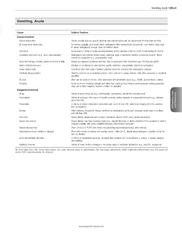
Vomiting, Acute 1293.e1


            Vomiting, Acute
  VetBooks.ir  Cause                             Salient Feature


            Gastrointestinal
              Dietary indiscretion               History; usually lack any specific physical exam abnormalities and not systemically ill other than vomiting
              GI foreign body obstruction        Sometimes palpable as GI mass effect; radiographs often demonstrate characteristic orad dilation (may need
                                                 to repeat radiographs); foreign object sometimes visible
              Parvovirus                         Acute onset of illness in poorly vaccinated/young animal; diarrhea common; point-of-care testing to confirm
              Functional obstruction (e.g., ileus, dysautonomia)  Radiographs demonstrate dilated bowel; additional signs of autonomic function sometimes present; history
                                                 sometimes suggestive (e.g., postoperative ileus)
              Acute hemorrhagic diarrhea syndrome (formerly HGE)  Always accompanied by bloody diarrhea; may be associated with Clostridium spp; PCV typically ≥60%
              Gastric dilatation/volvulus        Attempts to vomit may be unproductive; gastric distention; characteristic abdominal radiographs
              Linear foreign body                Cats more often than dogs; intestinal palpation abnormal; characteristic radiographic changes
              Intestinal intussusception         Diarrhea common as a precipitating factor; more common in young animals, often after parvovirus or intestinal
                                                 parasites
              GI ulcer                           Ulcer can be acute or chronic; often associated with identifiable cause (e.g., NSAID, glucocorticoid, uremia)
              Parasites                          Acute or chronic vomiting; typically well other than vomiting; fecal flotation and heartworm testing (especially
                                                 cats) can be false-negative; examine vomitus for parasites
            Nongastrointestinal
              Drugs                              History of recent drug use (e.g., methimazole, cyclosporine, amoxicillin-clavulanic acid)
              Intoxications                      History of exposure; other signs of toxicity common; testing depends on suspected intoxicant (e.g., ethylene
                                                 glycol test kits)                                                    Differentials, Lists,   and Mnemonics
              Pancreatitis                       ± History of dietary indiscretion; abdominal pain; point-of-care cPLI; abdominal imaging (US more sensitive
                                                 than radiographs)
              Uremia                             Other evidence of systemic illness; confirmed by biochemistry profile and urinalysis; acute onset more likely
                                                 with AKI than CKD
              Peritonitis                        Severe illness; abdominal pain common; abdominal effusion (FAST scan); abdominocentesis
              Sepsis (any source)                Severe illness; may have localizing signs (e.g., vaginal discharge or uterine distention from pyometra or painful
                                                 enlarged prostate with acute prostatitis/abscess); inflammatory leukogram
              Diabetic ketoacidosis              Often a history of PU/PD with newly recognized hyperglycemia/glucosuria; urine ketones
              Hypoadrenocorticism (Addison’s disease)  More often chronic or waxing and waning course; ± Na+↓K+↑; absent stress leukogram; baseline cortisol to
                                                 rule out disorder
              Acute hepatobiliary disorders      ± History of hepatotoxin exposure; increased liver enzymes (ALT predominant); ± icterus; ± hepatic imaging
                                                 abnormalities
              Vestibular disorder                History of travel (motion sickness) or neurologic signs of vestibular dysfunction (e.g., head tilt, nystagmus)
           AKI, acute kidney injury; CKD, chronic kidney disease; cPLI, canine pancreatic lipase; GI, gastrointestinal; HGE, hemorrhagic gastroenteritis; NSAID, nonsteroidal antiinflammatory drug; PCV, packed cell
           volume; PU/PD, polyuria/polydipsia; US, ultrasound.


                                                      www.ExpertConsult.com