
Page 1033 - Small Animal Internal Medicine, 6th Edition
P. 1033
CHAPTER 56 Clinical Conditions of the Dog and Tom 1005
be evaluated in azoospermic samples; a level of greater than
5000 IU/L suggests patency of the duct system and that a
VetBooks.ir complete ejaculate was obtained. Cytologic examination of
fine-needle aspirate of the testes (described earlier) can help
evaluate for the presence of spermatogenesis in azoospermic
dogs with low semen alkaline phosphatase by the identifica-
tion of spermatogonia, primary and secondary spermato-
cytes, spermatids, and spermatozoa; if normal, an obstructive
lesion is more likely. Prostatic evaluation is indicated as both
ductuli deferentes course through the prostate into the pros-
tatic urethra. Ultrasound of the reproductive tract should be
performed evaluating the prostate, epididymi, and urethra
for abnormalities that could interfere with semen outflow
(epididymal spermatocele, prostatitis).
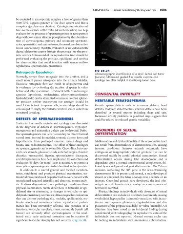
Retrograde Ejaculation FIG 56.34
Ultrasonographic identification of a small Sertoli cell tumor
Normally, semen flows antegrade into the urethra, and a (cursors). Ultrasound guided fine needle aspirate and
small amount passes retrograde into the urinary bladder. cytology are often helpful in identifying tumor type.
Excessive retrograde flow can result in oligospermia and
is confirmed by evaluating the number of sperm in urine
before and after ejaculation. Treatment with α-andrenergic
agonists (ephedrine, norfenefrine, phenylpropanolamine CONGENITAL INFERTILITY
hydrochloride) can be attempted to increase urethral sphinc-
ter pressure; neither testosterone nor estrogen should be HERITABLE TERATOSPERMIA
used. Urine is toxic to sperm cells, so stud dogs should be Genetic sperm defects such as acrosome defects, head
encouraged to empty their bladders before semen collection defects, midpiece abnormalities, and tail defects have been
or natural breeding. described in several species including dogs and cats.
Increased fertility problems in purebred dogs suggests this
DEFECTS OF SPERMATOGENESIS could be related to reduced genetic variability.
Testicular fine-needle aspirate and cytology can also assist
in the diagnosis of defects in spermatogenesis. Hyposper-
matogenesis and maturation defects can be detected. Defec- DISORDERS OF SEXUAL
tive spermatogenesis can occur secondary to direct thermal DIFFERENTIATION
scrotal insult (scrotal dermatitis), systemic disease, fever and
hyperthermia from prolonged exercise, certain drugs and Malformation and dysfunctionality of the reproductive tract
toxins, and endocrinopathies. The effect of these etiologies can result from abnormalities of chromosomal sex, causing
on spermatogenesis can be reversible. Cimetidine, ketocon- intersex conditions. Intersex animals commonly have
azole, sex steroids, glucocorticoids, anticholinergics, thiazide ambiguous or inappropriate external genitalia that can be
diuretics, propranolol, digoxin, spironolactone, diazepam, discovered readily by careful physical examination. Sexual
and chlorpromazine have been implicated. Re-collection and differentiation occurs during fetal development and is
evaluation 60 days (or more) later is necessary to permit a dependent upon a normal chromosomal complement, fol-
new cycle of spermatogenesis to be identified in the ejaculate. lowed by normal gonad and genitalia formation. The Y chro-
In addition to a careful general and focused (scrotum, mosome, containing the SRY gene, is the sex-determining
testes, epididymi, and prostate) physical examination, tes- chromosome. If it is present and normal, a male develops; if
ticular ultrasound should be performed in every patient with absent or abnormal, the fetus develops into a female or an
unexplained acquired infertility and abnormal semen analy- intersex. Once fetal gonads have developed, the fetal phe-
sis. It allows diagnosis of more pathologic conditions than notypic sexual characteristics develop as a consequence of
physical examination. Subtle differences in testicular or epi- hormones secreted.
didymal size or symmetry, or changes in testicular or epi- Physical findings in individuals with disorders of sexual
didymal consistency, warrant an ultrasonographic evaluation differentiation can include an os clitoris (commonly causing
that can disclose pathology (i.e., orchitis, epididymitis, tes- vestibulitis), hypospadias (commonly associated with incon-
ticular neoplasia) sometimes before reproductive perfor- tinence and exposure phimosis), cryptorchidism, and dis-
mance has been irreversibly affected. The presence of a placement of the prepuce (caudally) or vulva (cranially). Os
functional testicular neoplasia (most commonly Sertoli cell clitorises have been noted as an incidental finding during
tumor) can adversely affect spermatogenesis in the unaf- coxofemoral joint radiography; the reproductive status of the
fected testis; early unilateral castration can be curative if individuals was not reported. Normal estrous cycles can
significant testicular atrophy has not occurred (Fig. 56.34). be lacking in individuals with anomalous differentiation.