
Page 1028 - Small Animal Internal Medicine, 6th Edition
P. 1028
1000 PART VIII Reproductive System Disorders
TABLE 56.2
VetBooks.ir Bacterial Isolates From the Prepuce and Semen of
Tomcats With Normal Semen
PREPUCE SEMEN
n = 29 samples n = 29 samples
hemolytic E. coli hemolytic E. coli
Pseudomonas aeruginosa Pseudomonas aeruginosa
Proteus mirabilis Proteus mirabilis
Klebsiella oxytoca Klebsiella oxytoca
Streptococcus spp. Streptococcus spp.
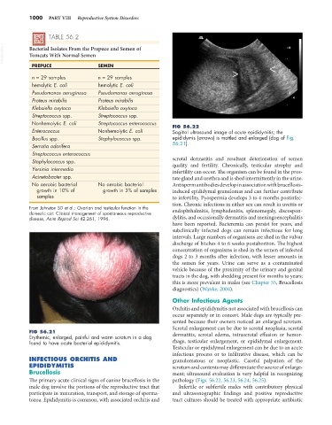
Nonhemolytic E. coli Streptococcus enterococcus FIG 56.22
Enterococcus Nonhemolytic E. coli Sagittal ultrasound image of acute epididymitis; the
Bacillus spp. Staphylococcus spp. epididymis (arrows) is mottled and enlarged (dog of Fig.
Serratia odorifera 56.21).
Streptococcus enterococcus
Staphylococcus spp. scrotal dermatitis and resultant deterioration of semen
quality and fertility. Chronically, testicular atrophy and
Yersinia intermedia infertility can occur. The organism can be found in the pros-
Acinetobacter spp. tate gland and urethra and is shed intermittently in the urine.
No aerobic bacterial No aerobic bacterial Antisperm antibodies develop in association with brucellosis-
growth in 10% of growth in 3% of samples induced epididymal granulomas and can further contribute
samples to infertility. Pyospermia develops 3 to 4 months postinfec-
tion. Chronic infections in either sex can result in uveitis or
From Johnston SD et al.: Ovarian and testicular function in the endophthalmitis, lymphadenitis, splenomegaly, discospon-
domestic cat: Clinical management of spontaneous reproductive
disease, Anim Reprod Sci 42:261, 1996. dylitis, and occasionally dermatitis and meningoencephalitis
have been reported. Bacteremia can persist for years, and
subclinically infected dogs can remain infectious for long
intervals. Large numbers of organisms are shed in the vulvar
discharge of bitches 4 to 6 weeks postabortion. The highest
concentration of organisms is shed in the semen of infected
dogs 2 to 3 months after infection, with lesser amounts in
the semen for years. Urine can serve as a contaminated
vehicle because of the proximity of the urinary and genital
tracts in the dog, with shedding present for months to years;
this is more prevalent in males (see Chapter 55, Brucellosis
diagnostics) (Wanke, 2004).
Other Infectious Agents
Orchitis and epididymitis not associated with brucellosis can
occur separately or in concert. Male dogs are typically pre-
sented because their owners noticed an enlarged scrotum.
Scrotal enlargement can be due to scrotal neoplasia, scrotal
FIG 56.21 dermatitis, scrotal edema, intrascrotal effusion or hemor-
Erythemic, enlarged, painful and warm scrotum in a dog
found to have acute bacterial epididymitis. rhage, testicular enlargement, or epididymal enlargement.
Testicular or epididymal enlargement can be due to an acute
infectious process or to infiltrative disease, which can be
INFECTIOUS ORCHITIS AND granulomatous or neoplastic. Careful palpation of the
EPIDIDYMITIS scrotum and contents may differentiate the source of enlarge-
Brucellosis ment; ultrasound evaluation is very helpful in recognizing
The primary acute clinical signs of canine brucellosis in the pathology (Figs. 56.22, 56.23, 56.24, 56.25).
male dog involve the portions of the reproductive tract that Infertile or subfertile males with contributory physical
participate in maturation, transport, and storage of sperma- and ultrasonographic findings and positive reproductive
tozoa. Epididymitis is common, with associated orchitis and tract cultures should be treated with appropriate antibiotic