
Page 1023 - Small Animal Internal Medicine, 6th Edition
P. 1023
CHAPTER 56 Clinical Conditions of the Dog and Tom 995
VetBooks.ir
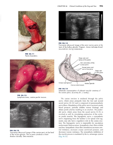
FIG 56.14
Transverse ultrasound image of the erect canine penis at the
level of the bulbus glandis. Priapism. Arrow indicates blood
accumulation during tumescence.
FIG 56.11
Penile laceration.
Deep vein and
artery of the penis
Vein and artery of the
bulbus penis
Dorsal artery and
vein of the penis
Deep vein
of the glans
Corpus spongiosum Bulbus glandis
Corpus cavernosum
FIG 56.15
Schematic representation of relevant vascular anatomy of
the canine penis. (Courtesy Dr. J. Lavely.)
FIG 56.12
Lymphosarcoma, canine penile mucosa.
The canine erection is mediated through the pelvic
nerve, which arises primarily from the first and second
0 sacral nerves (S1-S2) and is composed of parasympathetic
nerve fibers. Stimulation of the pelvic nerve increases penile
blood pressure, partially inhibits venous drainage, and
dilates penile arteries resulting in an erection. The puden-
dal nerve, which arises from the sacral nerves S1-S3, is
1
involved as well, by stimulating contraction of the extrin-
sic penile muscles. The hypogastric nerve, a sympathetic
nerve originating from the lumbar L1-L4 spinal cord seg-
ments, may also have a regulatory role in the canine erec-
2 tion. The hypogastric nerve is responsible for ejaculation
and prostatic fluid secretion. Sympathetic chain fibers inhibit
erection. Sympathetic chain fiber stimulation increases arte-
rial resistance, decreases corpus cavernosal pressure, and
FIG 56.13
Transverse ultrasound image of the canine penis at the level decreases venous resistance. The sympathetic inhibition of
of the bulbus glandis. The os penis produces a hard the erectile process is mediated by the α 1 -adrenergic system
shadow dorsally. Detumescence. (Fig. 56.15).