
Page 1025 - Small Animal Internal Medicine, 6th Edition
P. 1025
CHAPTER 56 Clinical Conditions of the Dog and Tom 997
located, very hyperechoic line. The epididymis (head, body, geriatric, a preoperative biochemical panel and urinalysis are
tail) is less echogenic than the testis. The ductus deferens is also reasonable. Hyperestrogenism can cause atrophy of the
VetBooks.ir difficult to visualize. The spermatic cord is adjacent to the unaffected testis resulting in azoospermia, which can be
noted clinically before a testicular mass is diagnosed.
head of the epididymis and has obvious, tortuous, small
After castration, histopathology of the testicular mass
diameter veins. Testicular neoplasms appear as variably cir-
cumscribed masses, hypo- to hyperechoic, which may with evaluation of the local lymphatics is indicated. Most
obscure the mediastinum testis (Fig. 56.18). The appearance testicular neoplasia in the dog is cured by castration as the
is not specific for tumor type; masses tend to change from potential for distant metastasis is low; local metastasis (intra-
hypoechoic to mixed echogenicity with growth likely due to abdominal via regional lymphatics) tends to occur late with
necrosis and hemorrhage. Testicular tumors commonly chronicity.
cause enlargement of the testis with chronicity; some cause
paraneoplastic syndromes.
In dogs, Sertoli cell tumors, Leydig cell (interstitial cell) MICROBIOLOGY AND MALE FERTILITY
tumors, and seminomas occur with about equal frequency
in scrotal testes; intraabdominal testicular neoplasia is most Previously fertile stud dogs producing small litter size or
commonly the Sertoli cell tumor. Sertoli cell and interstitial failing to impregnate normal bitches with good husbandry
(Leydig) cell tumors can produce hormones, particularly and normal breeding behavior should have semen evaluation
estrogen, which can cause paraneoplastic syndromes. performed (see semen collection, Chapter 54). If the semen
Although these tumors usually are clinically silent, the pro- is abnormal and associated with inflammatory cells or pain
duction of estrogen, progesterone, and corticosteroids has during ejaculation, it should be submitted for aerobic, anaer-
also been described. Estrogen excess and feminizing syn- obic, and Mycoplasma spp. culture, and B. canis testing
dromes may occur from the peripheral aromatization of tes- should always be performed (see Chapter 54). Semen is
tosterone or from the direct production of estradiol by the judged to be abnormal if no semen (aspermia), no sperm
tumor itself. These include atrophy of the contralateral testis, (azoospermia), or inadequate numbers of sperm (<200-400
bone marrow suppression, pendulous prepuce, gynecomas- million plus per ejaculate [oligospermia]) are present; if
tia, alopecia, hyperpigmentation, and squamous metaplasia sperm motility is less than 75% to 90% progressively motile
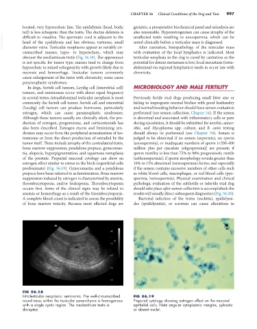
of the prostate. Preputial mucosal cytology can show an (asthenospermia); if sperm morphology reveals greater than
estrogen effect similar to estrus in the bitch (superficial cells 10% to 15% abnormal (teratospermia) forms; and especially
predominate) (Fig. 56.19). Gynecomastia and a pendulous if the semen contains excessive numbers of other cells such
prepuce have been referred to as feminization. Bone marrow as white blood cells, macrophages, or red blood cells (pyo-
suppression induced by estrogen is characterized by anemia, spermia, hemospermia). Physical examination and clinical
thrombocytopenia, and/or leukopenia. Thrombocytopenia pathologic evaluation of the subfertile or infertile stud dog
occurs first. Some of the clinical signs may be related to should take place after semen collection is accomplished; the
anemia or hemorrhage as a result of the thrombocytopenia. results will usually direct subsequent diagnostics (Fig. 56.20).
A complete blood count is indicated to assess the possibility Bacterial infection of the testes (orchitis), epididymi-
of bone marrow toxicity. Because most affected dogs are des (epididymitis), or scrotum can cause alterations in
FIG 56.18
Intratesticular neoplasia: seminoma. The well-circumscribed FIG 56.19
round mass within the testicular parenchyma is homogenous Preputial cytology showing estrogen effect on the mucosal
with a single cystic region. The mediastinum testis is epithelial cells. Note angular cytoplasmic margins, pyknotic
disrupted. or absent nuclei.