
Page 1021 - Small Animal Internal Medicine, 6th Edition
P. 1021
CHAPTER 56 Clinical Conditions of the Dog and Tom 993
VetBooks.ir
A A
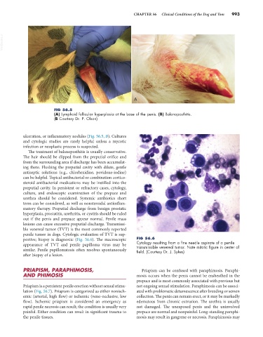
FIG 56.5
(A) Lymphoid follicular hyperplasia at the base of the penis. (B) Balanoposthitis.
(B Courtesy Dr. P. Olson)
ulceration, or inflammatory nodules (Fig. 56.5, B). Cultures
and cytologic studies are rarely helpful unless a mycotic
infection or neoplastic process is suspected.
The treatment of balanoposthitis is usually conservative.
The hair should be clipped from the preputial orifice and
from the surrounding area if discharge has been accumulat-
ing there. Flushing the preputial cavity with dilute, gentle
antiseptic solutions (e.g., chlorhexidine, povidone-iodine)
can be helpful. Topical antibacterial or combination cortico-
steroid antibacterial medications may be instilled into the
preputial cavity. In persistent or refractory cases, cytology,
culture, and endoscopic examination of the prepuce and
urethra should be considered. Systemic antibiotics short
term can be considered, as well as nonsteroidal antiinflam-
matory therapy. Preputial discharge from benign prostatic
hyperplasia, prostatitis, urethritis, or cystitis should be ruled
out if the penis and prepuce appear normal. Penile mass
lesions can cause excessive preputial discharge. Transmissi-
ble venereal tumor (TVT) is the most commonly reported
penile tumor in dogs. Cytologic evaluation of TVT is sup-
portive; biopsy is diagnostic (Fig. 56.6). The macroscopic FIG 56.6
appearance of TVT and penile papilloma virus may be Cytology resulting from a fine needle aspirate of a penile
transmissible venereal tumor. Note mitotic figure in center of
similar. Penile papillomatosis often resolves spontaneously field. (Courtesy Dr. J. Sykes)
after biopsy of a lesion.
PRIAPISM, PARAPHIMOSIS, Priapism can be confused with paraphimosis. Paraphi-
AND PHIMOSIS mosis occurs when the penis cannot be ensheathed in the
prepuce and is most commonly associated with previous but
Priapism is a persistent penile erection without sexual stimu- not ongoing sexual stimulation. Paraphimosis can be associ-
lation (Fig. 56.7). Priapism is categorized as either nonisch- ated with problematic detumescence after breeding or semen
emic (arterial, high flow) or ischemic (veno-occlusive, low collection. The penis can remain erect, or it may be markedly
flow). Ischemic priapism is considered an emergency as edematous from chronic extrusion. The urethra is usually
rapid penile necrosis can result; the condition is usually very not damaged. The unexposed penis and the uninvolved
painful. Either condition can result in significant trauma to prepuce are normal and nonpainful. Long-standing paraphi-
the penile tissues. mosis may result in gangrene or necrosis. Paraphimosis may