
Page 1020 - Small Animal Internal Medicine, 6th Edition
P. 1020
992 PART VIII Reproductive System Disorders
PERSISTENT PENILE FRENULUM scrotum. Physical examination of a dog with abnormal
semen should always include close visual evaluation of the
VetBooks.ir Under the influence of androgens, the surfaces of the glans ventral scrotum. Scrotal mast cell tumors can incite local
inflammation.
penis and the preputial mucosa normally separate before or
Appropriate topical and systemic therapies should be
within weeks of birth. If this separation does not occur, con-
nective tissue persists between the penis and the prepuce. In instituted, and prevention of excoriation by the use of Eliza-
dogs the persistent penile frenulum is usually located on the bethan collars encouraged. Nonsteroidal antiinflammatory
ventral midline of the penis. A persistent penile frenulum drugs such as carprofen (Rimadyl® Pfizer), meloxicam
may cause no clinical signs, or it may be associated with (Metacam® Boehringer Ingelheim), firocoxib (Previcox®
preputial discharge or excessive licking of the prepuce. Per- Merial), or grapiprant (Galliprant® Aratana) are useful. Nar-
sistent frenulum may cause the penis to deviate ventrally or cotics (Tramadol) may be necessary short term for adequate
laterally so that the dog or tomcat is unable or unwilling to analgesia. Broad spectrum antibiotics appropriate for pyo-
mate, or it may interfere with normal tumescence (Fig. 56.3). derma, such as cephalexin or cefpodoxime proxetil (Simpli-
The diagnosis is made by visual examination. Treatment is cef® Pfizer), are appropriate. The use of corticosteroids should
surgical excision, which can often be done using just seda- be avoided. Normalization of spermatogenesis can take more
tion with local anesthesia as the frenulum tends to be a sheer, than 60 days.
avascular membrane.
BALANOPOSTHITIS
URETHRAL PROLAPSE
Inflammation or infection of the preputial cavity and penis,
Urethral prolapse occurs most commonly in Bulldogs, balanoposthitis, is common in dogs and rare in cats. Normal
French Bulldogs, and Boston Terriers, and is likely familial. scant white smegma in an intact male dog should not be
Eversion of the urethral mucosa at the distal tip of the penis mistaken for balanoposthitis. The causative organisms are
results in refractory hemorrhage. The condition may be asso- usually members of the normal preputial flora, although
ciated with the increased intraabdominal pressure associated overgrowth of one organism or a predominance of Pseudo-
with the brachycephalic syndrome. Surgical revision is indi- monas spp can occur. Balanoposthitis usually causes no clini-
cated as the condition will not resolve spontaneously. Pre- cal signs other than a purulent preputial discharge that varies
venting erection during recovery is important; breeding from mucoid to copious green pus accompanied by excessive
these dogs can cause relapse, and castration should be sug- licking. The discharge associated with balanoposthitis is not
gested for both therapeutic and ethical reasons. sanguineous unless the cause is neoplasia or accumulated
foreign material. Lymphoid follicular hyperplasia is com-
monly also present and thought to develop as a result of
SCROTAL DERMATITIS chronic irritation (Fig. 56.5, A).
The diagnosis of balanoposthitis is made by physical
Scrotal dermatitis can result from trauma, contact irritants examination of the penis and preputial cavity all the way to
or hypersensitivities, excessively warm bedding/heating the fornix, in a search for foreign material, neoplasia,
pads, burns, frostbite, envenomation, shaving, allergic der-
matopathies, or intrascrotal pathology inciting excoriation
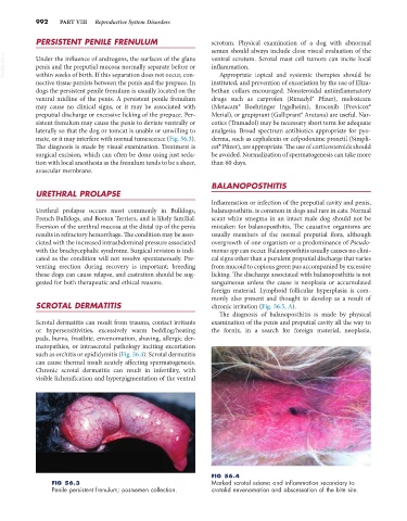
such as orchitis or epididymitis (Fig. 56.4). Scrotal dermatitis
can cause thermal insult acutely affecting spermatogenesis.
Chronic scrotal dermatitis can result in infertility, with
visible lichenification and hyperpigmentation of the ventral
FIG 56.4
FIG 56.3 Marked scrotal edema and inflammation secondary to
Penile persistent frenulum; postsemen collection. crotalid envenomation and abscessation of the bite site.