
Page 129 - Small Animal Internal Medicine, 6th Edition
P. 129
CHAPTER 5 Congenital Cardiac Disease 101
MURMUR
VetBooks.ir Consider history and other physical
and CV exam findings
Evaluate pulse, precordium, radiographs,
ECG, echocardiogram, and PCV
Normal findings Abnormal findings
Systolic murmur Systolic murmur Both systolic +
r/o: diastolic murmurs
Innocent murmur
Physiologic murmur
(e.g., fever, anemia)
Mild congenital defect Animal
acyanotic Loud at time of S 2
(continuous)
r/o:
PDA
Heard best on: Animal
cyanotic
r/o:
Left Right T of F Soft at time of S 2
(“to and fro”)
hemithorax hemithorax Pulmonary r/o:
r/o: r/o: hypertension SAS + aortic
PS (base) VSD (reversed PDA, insufficiency
ASD (base) ECD VSD, or VSD + aortic
Acyanotic T of F T dysplasia ASD) insufficiency
(base) SAS Complex anomaly
SAS (3rd-4th ICS)
M dysplasia (apex)
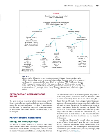
FIG 5.1
Flow chart for differentiating murmurs in puppies and kittens. Thoracic radiographs,
±ECG, also can help screen for structural abnormalities; however, referral to a veterinary
cardiologist is recommended for more definitive evaluation. ASD, Atrial septal defect;
ECD, endocardial cushion defect; ECG, electrocardiogram; ICS, intercostal space; M,
mitral valve; PCV, packed cell volume; PDA, patent ductus arteriosus; r/o, rule out; SAS,
subaortic stenosis; T, tricuspid valve; T of F, tetralogy of Fallot; VSD, ventricular septal
defect.
EXTRACARDIAC ARTERIOVENOUS and contains less smooth muscle and a greater proportion of
SHUNT elastic fibers, similar to the aortic wall. It is therefore unable
to constrict effectively. When the ductus fails to close, blood
The most common congenital arteriovenous shunt is PDA. shunts through it from the descending aorta into the pulmo-
Rarely, similar hemodynamic and clinical abnormalities are nary artery. Because aortic pressure normally is higher than
caused by an aorticopulmonary window (a communica- pulmonic pressure throughout the cardiac cycle, shunting
tion between the ascending aorta and pulmonary artery) occurs continuously during both systole and diastole. This
or some other functionally similar communication in the left-to-right shunt causes a volume overload of the pulmo-
hilar region. nary circulation, left atrium (LA), and left ventricle (LV). The
shunt volume is directly related to the pressure difference
(gradient) between the two circulations and the diameter
PATENT DUCTUS ARTERIOSUS of the ductus.
Hyperkinetic (“bounding”) arterial pulses are charac-
Etiology and Pathophysiology teristic of PDA. Blood runoff from the aorta into the pul-
The ductus normally constricts to become functionally monary system allows diastolic aortic pressure to decrease
closed within hours of birth. Structural changes and perma- rapidly below normal. The widened pulse pressure (differ-
nent closure occur over the ensuing weeks. The ductal wall ence between systolic and diastolic pressure) causes palpably
in animals with an inherited PDA is histologically abnormal stronger arterial pulses (Fig. 5.2).