
Page 131 - Small Animal Internal Medicine, 6th Edition
P. 131
CHAPTER 5 Congenital Cardiac Disease 103
pulmonary artery pressure rises toward aortic pressure, pro- Diagnosis
gressively less blood shunting occurs. If pulmonary artery Radiographs usually show cardiac elongation (left heart dila-
VetBooks.ir pressure exceeds aortic pressure, shunt reversal (right-to-left tion), left atrial (LA) and auricular enlargement, and pulmo-
nary overcirculation (Table 5.2). A bulge often is evident in
flow) occurs. Approximately 15% of dogs with PDA have
the descending aorta (“ductus bump”), main pulmonary
reversed (right-to-left) shunting. However, as most such
shunts are already “reversed” (right-to-left) by the time of trunk, or both (Fig. 5.3). The triad of all three bulges (i.e.,
first evaluation, it is difficult to know whether these patients pulmonary trunk, aorta, and left auricle), located in that
have retained fetal pulmonary vascular resistance (congenital order from the 1 to 3 o’clock position on a dorsoventral (DV)
pulmonary hypertension) causing right-to-left shunting radiograph, is a classic finding but not always seen. Animals
from birth or whether shunt flow actually reversed postna- with left-sided CHF also show evidence of pulmonary
tally following pulmonary vascular changes from volume edema. Characteristic ECG findings suggest LV and LA
overload. enlargement, including wide P waves, tall R waves, and often
deep Q waves in leads II, aVF, and CV 6 LL. Changes in the
Clinical Features ST-T segment secondary to LV enlargement can occur.
The left-to-right shunting PDA is by far the most common However, the ECG is normal in some animals with PDA.
form; clinical features of reversed PDA are described on page Most patients have normal sinus rhythm, although ventricu-
115. The prevalence of PDA is higher in certain breeds of lar or supraventricular arrhythmias (including atrial fibrilla-
dogs; a polygenic inheritance pattern is thought to exist, tion) can occur.
particularly in miniature Poodles. The prevalence is two or Echocardiography also shows left heart enlargement and
more times greater in female than male dogs. Most animals pulmonary trunk dilation. LV fractional shortening can be
are asymptomatic when first diagnosed, although some normal or decreased, and the E point–septal separation is
patients may present with clinical signs of left-sided CHF often increased. The ductus itself can be difficult to visualize
including exercise intolerance, tachypnea, or cough. A con- because of its location between the descending aorta and
tinuous murmur heard best high at the left base (see p. 11), pulmonary artery; angulation from the left cranial short axis
often with a precordial thrill, is typical for a left-to-right view usually is most helpful. Doppler interrogation docu-
PDA; sometimes only the systolic component of the murmur ments continuous, turbulent flow into the pulmonary artery
is heard more caudally near the mitral valve area. Other (Fig. 5.4). The maximum aortic-to-pulmonary artery pres-
findings include hyperkinetic (bounding, “water hammer”) sure gradient can be estimated using velocity of systolic PDA
arterial pulses and pink mucous membranes. flow. Cardiac catheterization generally is unnecessary for
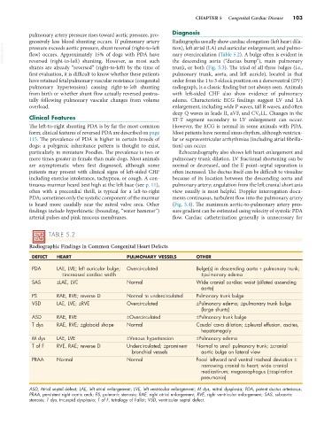
TABLE 5.2
Radiographic Findings in Common Congenital Heart Defects
DEFECT HEART PULMONARY VESSELS OTHER
PDA LAE, LVE; left auricular bulge; Overcirculated Bulge(s) in descending aorta + pulmonary trunk;
±increased cardiac width ±pulmonary edema
SAS ±LAE, LVE Normal Wide cranial cardiac waist (dilated ascending
aorta)
PS RAE, RVE; reverse D Normal to undercirculated Pulmonary trunk bulge
VSD LAE, LVE; ±RVE Overcirculated ±Pulmonary edema; ±pulmonary trunk bulge
(large shunts)
ASD RAE, RVE ±Overcirculated ±Pulmonary trunk bulge
T dys RAE, RVE; ±globoid shape Normal Caudal cava dilation; ±pleural effusion, ascites,
hepatomegaly
M dys LAE, LVE ±Venous hypertension ±Pulmonary edema
T of F RVE, RAE; reverse D Undercirculated; ±prominent Normal to small pulmonary trunk; ±cranial
bronchial vessels aortic bulge on lateral view
PRAA Normal Normal Focal leftward and ventral tracheal deviation ±
narrowing cranial to heart; wide cranial
mediastinum; megaesophagus (±aspiration
pneumonia)
ASD, Atrial septal defect; LAE, left atrial enlargement; LVE, left ventricular enlargement; M dys, mitral dysplasia; PDA, patent ductus arteriosus;
PRAA, persistent right aortic arch; PS, pulmonic stenosis; RAE, right atrial enlargement; RVE, right ventricular enlargement; SAS, subaortic
stenosis; T dys, tricuspid dysplasia; T of F, tetralogy of Fallot; VSD, ventricular septal defect.